The Changes of Sagittal Alignment after Anterior Interbody Fusion with Posterior Fixation in Spondylolisthesis of the Lumbar Spine
- Affiliations
-
- 1Department of Orthopaedic Surgery, Ajou University School of Medicine, Korea.
- 2Department of Orthopaedic Surgery, Konyang University School of Medicine, Korea. spinekyc@kyuh.co.kr
- KMID: 1839983
- DOI: http://doi.org/10.4184/jkss.2004.11.3.131
Abstract
- STUDY DESIGN: A prospective radiological assessment was conducted.
OBJECTIVES
To analyze the changes in the height of the intervertebral disc, the slippage, slip angle, lumbar lordotic angle and sacral inclination after anterior lumbar interbody fusion and posterior pedicle screw fixation in a lumbar spondylolisthesis. SUMMARY OF LITERATURE REVIEW: The anterior lumbar interbody fusion causes changes in the lumbar sagittal alignment.
METHODS
The mini-open anterior lumbar interbody fusion and pedicle screw fixation was undertaken in 33 cases from April 1995 to November 2003. MRI was done before and 6 months after surgery. The measuring factors were the heights of the intervertebral disc, slippage, slip angle, lumbar lordotic angle and sacral inclination. The measuring factors were independently assessed three times by three different orthopedic surgeons. The postoperative changes in measuring the factors were analyzed by a paired t-test statistically.
RESULTS
The height of the intervertebral disc was increased by a mean of 14.0%, slippage was reduced by a mean of 2.8%, the slip angle was reduced by a mean of 16.0%, the lumbar lordotic angle was increased by a mean of 15.6% and the scaral inclination was increased by a mean of 3.0%. There was significance in the increase in the disc height, the reduction of slippage and the slip angle, and the increase in lumbar lordotic angle, but there were no significance regarding the changes in sacral inclina-tion.
CONCLUSIONS
The anterior lumbar interbody fusion and the pedicle screw fixation significantly improved the height of the intervertebral disc, slippage, slip angle, and lumbar lordotic angle, except sacral inclination.
Keyword
MeSH Terms
Figure
-

Fig. 1. Radiological measurement of disc height.
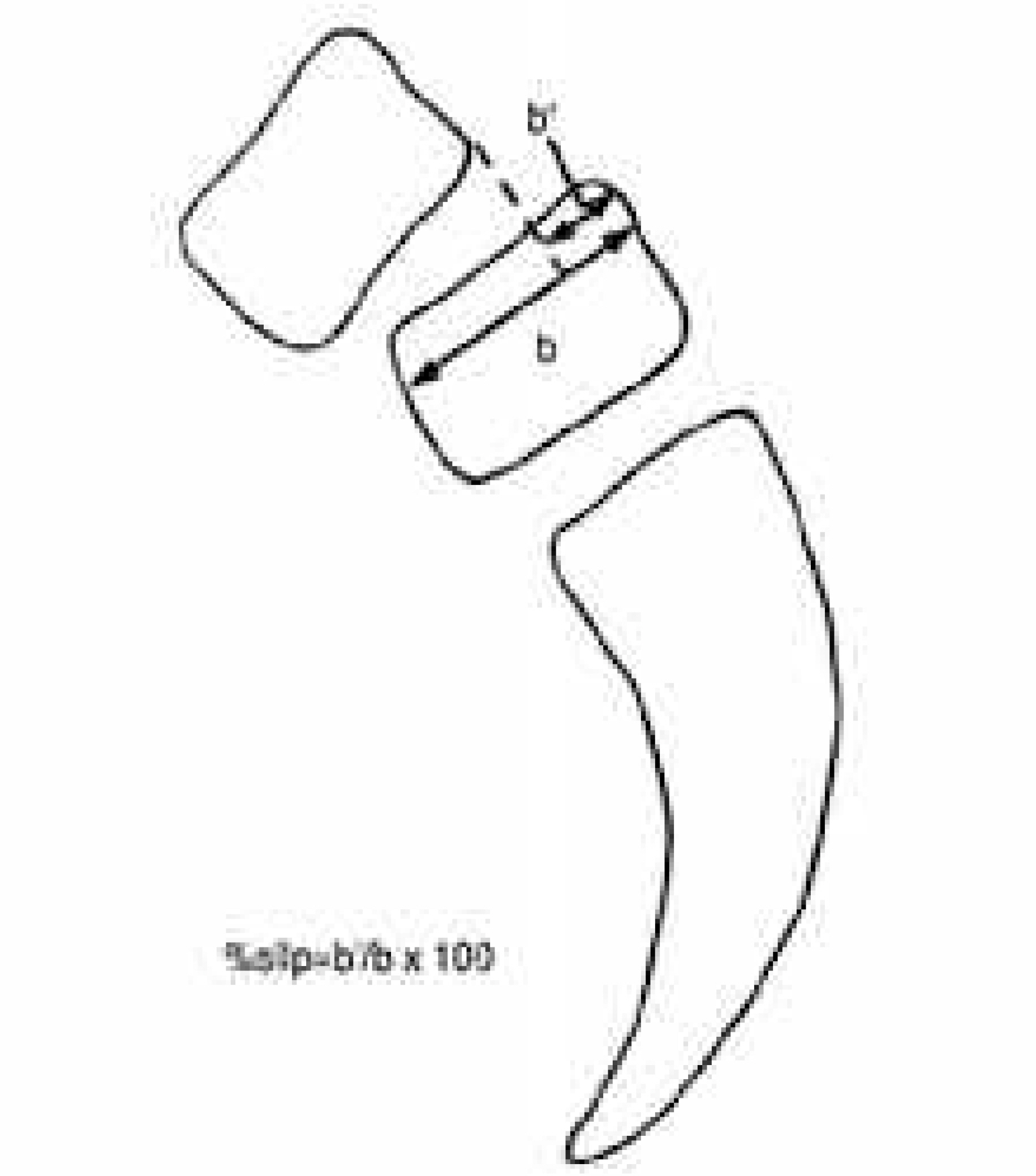
Fig. 2. Radiological measurment of slip percentage.

Fig. 3. Radiological measurment of slip angle (c).

Fig. 4. Radiological measurment of lumbar lordotic angle(d) and sacral inclination(e).
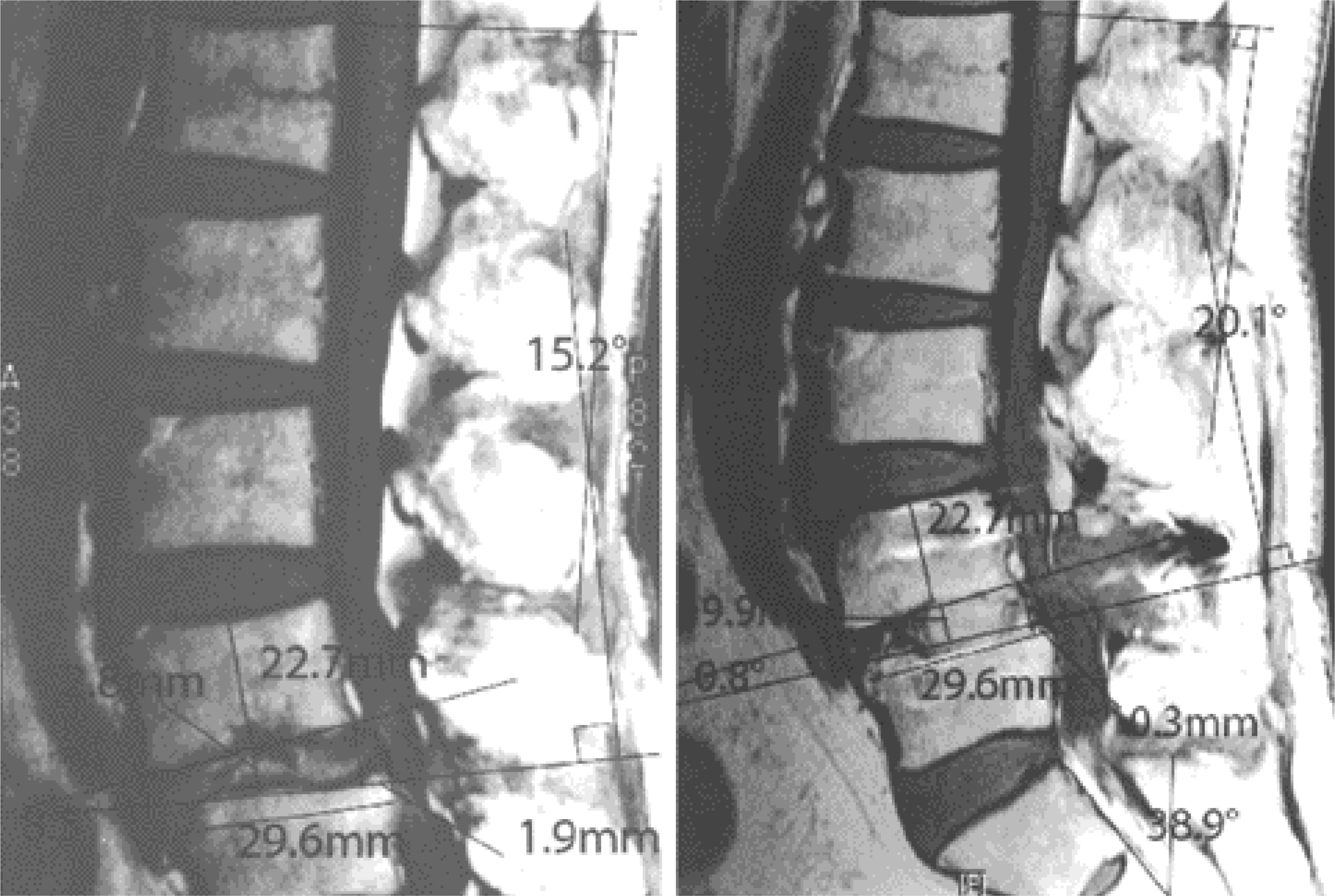
Fig. 5. A 61-year-old woman with degenerative spondylolisthesis L4 on L5. (A) sagittal image of level of the midportion of vertebral body on preoperative MRI shows 12.4% in disc height rate, 3.4% in slip, 8.5° in slip angle, 15.2° in lumbar lordotic angle,42.4° in sacral inclination. (B) sagittal image of level of the midportion of vertebral body on postoperative MRI shows 47.1% in disc height rate, 1.2% in slip, 0.8° in slip angle, 20.1° in lumbar lordotic angle, 38.9° in sacral inclination.

Fig. 6. A 48-year-old woman with isthmic spondylolisthesis L4 on L5. (A) sagittal image of level of the midportion of vertebral body on preoperative MRI shows 19.1% in disc height rate, 3% in slip, 0.8° in slip angle, 36.2° in lumbar lordotic angle, 44.0° in sacral inclination. (B) sagittal image of level of the midportion of vertebral body on postoperative MRI shows 36.1% in disc height rate, 1.3% in slip, 1.9° in slip angle, 32.4° in lumbar lordotic angle, 36.5° in sacral inclination.
Reference
-
1). Carragee EJ. Single-level posterolateral arthrodesis, with or without posterior decompression, for the treatment of isthmic spondylolisthesis in adults. A prospective, randomized study. J Bone Joint Surg. 1997; 79-A:1175–1180.
Article2). De Loubress CG, Bon T, Deburge A, Lassale B, Benoit M. Posterolateral fusion for radicular pain in isthmic spondylolisthesis. Clin Orthop. 1996; 323:194–201.3). Deguchi M, Rapoff AJ, Zdeblick TA. Poste rola te ral fusion for isthmic spondylolisthesis in adults: analysis of fusion rate and clinical results. J Spinal Disord. 1998; 11(6):459–464.4). Freeman BJC, Licina P, Mehdian SH. Posterior lumbar interbody fusion combined with instrumented posterolateral fusion: 5-year results in 60 patients. Eur Spine J. 2000; 9:42–46.
Article5). Kim EH, Woo BC, Koh ES, Cho DY. The change of segmental sagittal angle in low-grade spondylolisthesis after pedicle screw fixation with or without PLIF-PLIF+PLF versus PLF group. J Kor Orthop Assoc. 1997; 32(4):1098–1106.6). Enker P, Steffee AD. Interbody fusion and instrumentation. Clin Orthop. 1994; 300:90–101.
Article7). Suk KS, Jeon CH, LEE HM, Kim NH, Kim HC. Comparison between posterolateral fusion with pedicle screw fixation and anterior interbody fusion with pedicle screw fixation in spondylolytic spondylolisthesis of the lumbar spine, J Kor Spine Surg. 1999; 6(3):397–406.8). Suk SI, Lee CK, Kim WJ, Lee JH, Cho KJ, Kim HG. Adding posterior lumbar interbody fusion to pedicle screw fixation and posterolateral fusion after decompression in spondylolitic spondylolisthesis. Spine. 1997; 22:210–219.9). Wiltse LL, Winter RB. Terminology and measurement of spondylolisthesis. J Bone Joint Surg. 1983; 65-A:768–772.
Article10). Schnee CL, Freese A, Ansell LV. Outcome analysis for adults with spodylolisthesis treatment with posterolateral fusion and transpedicular screw fixation. J Neurosurg. 1997; 86(1):56–63.11). Vaccaro AR, Martyak GG, Madigan L. Adult isthmic spondylolisthesis. Orthopedics. 2001; 24:1172–1177.
Article12). Kim KT, Suk KS, Kim JM, Sin DJ, Shin NC. Results of the posterolateral fusion using a pedicle screw for the spondylolisthesis. J Kor Orthop Assoc. 2001; 36(2):167–172.
Article13). Inoue S, Watanabe T, Sumio G, Takahashi K, Takada K, Sho E. Degenerative spondylolisthesis. Pathophysiolo -gy and results of anterior interbody fusion fusion. Clin Orthop. 1988; 227:90–98.14). Ishihara H, Osada R, Kanamori M, et al. Minimum 10-year followup study of anterior lumbar interbody fusion for isthmic spondylolisthesis. J Spinal Disord. 2001; 14(2):91–99.
Article15). Jahng JH, Baek DH, Chang H, et al. Anterior interbody fusion and posterior instrumentation for degenerative lumbar spondylolisthesis. J Kor Orthop Assoc. 1988; 33(2):359–366.16). Madan SS, Boeree NR. Comparison of instrumented anterior interbody fusion with instrumented circumferential lumbar fusion. Eur Spine J. 2003; 12(6):567–575.
Article17). Jeon CH. Anterior interbody fsion for spondylolisthesis. J Kor Spine Surg. 2001; 8(3):350–355.18). Kim YT, Lee CS, Na HY, Lee CW. A comparison of surgical treatment in isthmic and degenerative spondylolisthesis. J Kor Orthop Assoc. 1998; 33(7):1672–1634.
Article19). Lee KY, Sohn SK, Kim SW, Lee SW. Outcome of two fusion methods in isthmic and degenerative spondylolisthesis of the lumbar spinen. J Kor Spine Surg. 2002; 9(4):313–321.20). Rosenberg NJ. Degenerative spondylolisthesis: Predis -posing factors. J Bone Joint Surg. 1975; 57-A:467–474.21). Lowe RW, Hayes TD, Kaye J, Bagg RJ, Luekens CA. Standing roentgenograms in spondylolisthesis. Clin Orthop. 1976; 117:80–84.
Article22). Saraste H, Brorstrom L-A, Aparisi T, Axdorph G. Radiographic measurement of the lumbar spine: A clinical and experimental study in man. Spine. 1985; 10:236–241.
- Full Text Links
-
- Actions
-
Cited
- CITED
-
- Close
- Share
-
- Similar articles
-
- The Change of Segmental Sagittal angle in Low - grade spondylolisthesis after Pedicular Screw Fixation with or without PLIF - PLIF + PLF versus PLF groups -
- The Changes in the Dimensions of Neural Foramen After Anterior Interbody Fusion in the Spondylolisthesis
- Comparison between Posterolateral Fusion with Pedicle Screw Fixation and Anterior Interbody Fusion with Pedicle Screw Fixation in Spondylolytic Spondylolisthesis of the Lumbar Spine
- Height Changes of Intervertebral Disc and Neural Foramen after Anterior Lumbar Interbody Fusion in the Lumbar Spine
- Effect of the Pedicle Screw Fixation on the Anterior Lumbar Interbody Fusion Using the Freeze - Dried Structural Allograft
