Korean J Urogenit Tract Infect Inflamm.
2015 Apr;10(1):49-52. 10.14777/kjutii.2015.10.1.49.
Encrusted Cystitis and Pyeloureteritis in Patient with Hepatocellular Carcinoma
- Affiliations
-
- 1Department of Urology, Wonkwang University School of Medicine and Hospital, Iksan, Korea. sc.park@wonkwang.ac.kr
- 2Institute of Wonkwang Medical Science, Wonkwang University School of Medicine and Hospital, Iksan, Korea.
- 3Department of Radiology, Wonkwang University School of Medicine and Hospital, Iksan, Korea.
- 4Hana Clinic, Naju, Korea.
- KMID: 1839389
- DOI: http://doi.org/10.14777/kjutii.2015.10.1.49
Abstract
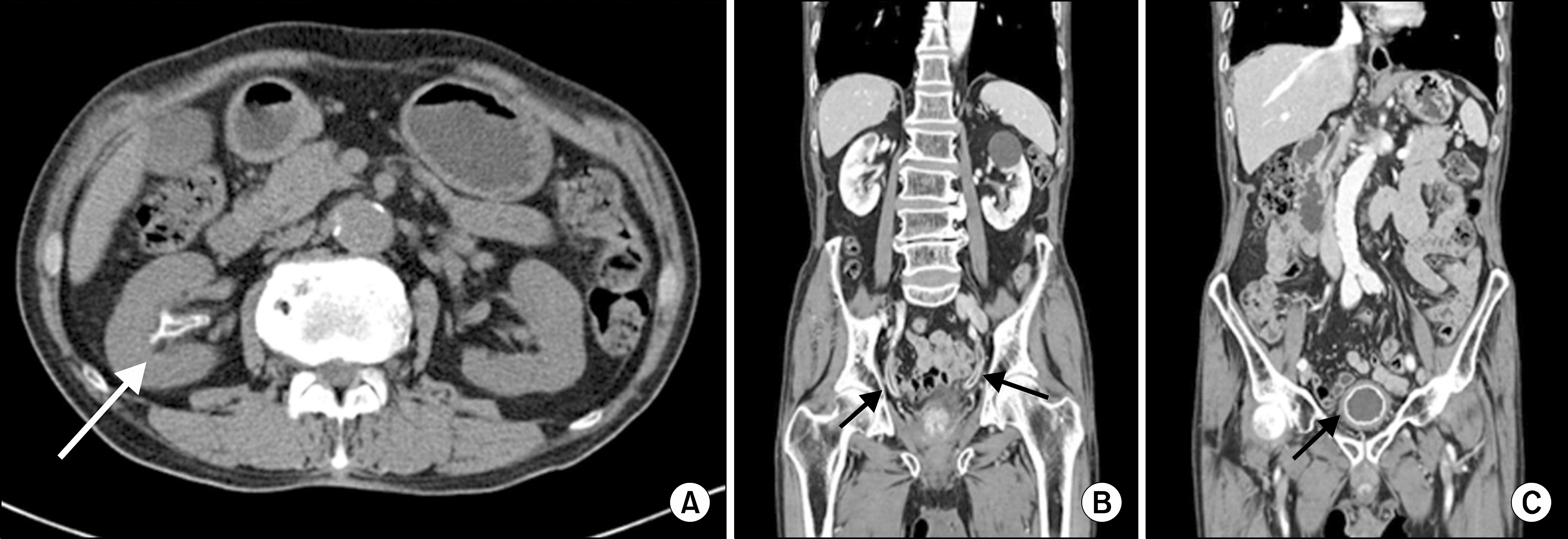
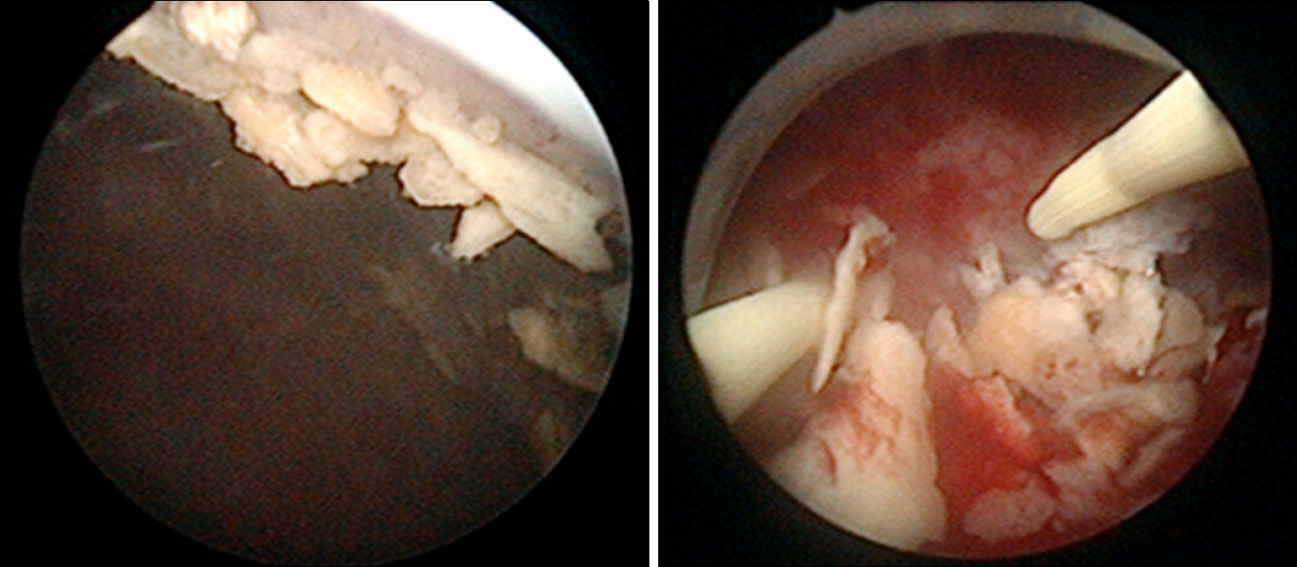
- Encrusted cystitis and pyeloureteritis are rare chronic infectious conditions characterized by mucosal inflammation and encrustations of the urinary tract. It is caused by fastidious growing urea splitting microorganisms, mainly Corynebacterium. Herein, we report an unusual case of an 80-year-old man with encrusted cystitis and pyeloureteritis who was previously treated with transcatheteral arterial chemoembolization for hepatocellular carcinoma. Abdomino-pelvic computerized tomography showed a bilateral hydronephrosis with calcifications of renal pelvis, ureter, and bladder. Cystoscopy showed calcified bladder mucosa with necrosis and bleeding. After transurethral removal of calcified plaques, the patient was treated with antibiotic and oral urine acidification. One-month follow-up cystoscopy showed that inflammation was improved and calcification was significantly reduced.
Keyword
MeSH Terms
Figure
Reference
-
1. Hara K, Kohno S, Koga H, Kaku M, Tomono K, Sakata S, et al. Infections in patients with liver cirrhosis and hepatocellular carcinoma. Intern Med. 1995; 34:491–5.
Article2. Tanaka T, Yamashita S, Mitsuzuka K, Yamada S, Kaiho Y, Nakagawa H, et al. Encrusted cystitis causing postrenal failure. J Infect Chemother. 2013; 19:1193–5.
Article3. Lieten S, Schelfaut D, Wissing KM, Geers C, Tielemans C. Alkaline-encrusted pyelitis and cystitis: an easily missed and life-threatening urinary infection. BMJ Case Rep. 2011; 2011.
Article4. Pomakova D, Segal BH. Prevention of infection in cancer patients. Cancer Treat Res. 2014; 161:485–511.
Article5. Bodey GP, Buckley M, Sathe YS, Freireich EJ. Quantitative relationships between circulating leukocytes and infection in patients with acute leukemia. Ann Intern Med. 1966; 64:328–40.
Article6. Soriano F, Tauch A. Microbiological and clinical features of Corynebacterium urealyticum: urinary tract stones and genomics as the Rosetta Stone. Clin Microbiol Infect. 2008; 14:632–43.
Article7. Meria P, Desgrippes A, Arfi C, Le Duc A. Encrusted cystitis and pyelitis. J Urol. 1998; 160:3–9.
Article8. Penta M, Fioriti D, Chinazzi A, Pietropaolo V, Conte MP, Schippa S, et al. Encrusted cystitis in an immunocompromised patient: possible coinfection by Corynebacterium urealyticum and E. coli. Int J Immunopathol Pharmacol. 2006; 19:241–4.9. Eschwege P, Hauk M, Blanchet P, Hiesse C, Vieillefond A, Blery M, et al. Imaging analysis of encrusted cystitis and pyelitis in renal transplantation. Transplant Proc. 1995; 27:2444–5.10. Carlsson S, Wiklund NP, Engstrand L, Weitzberg E, Lundberg JO. Effects of pH, nitrite, and ascorbic acid on nonenzymatic nitric oxide generation and bacterial growth in urine. Nitric Oxide. 2001; 5:580–6.
Article
- Full Text Links
- Actions
-
Cited
- CITED
-
- Close
- Share
- Similar articles
-
- A case of eosinophilic cystitis
- Metastatic Omental Hepatocellular Carcinoma: Two Cases Report
- Two Cases of Cutaneous Metastasis Originating from Hepatocellular Carcinoma
- Hepatocellular Carcinoma Arising in Hepatocellular Adenoma
- A Case of Cutaneous Acrometastasis of Hepatocellular Carcinoma to the Finger


