A case of papillary squamotransitional cell carcinoma in the vagina
- Affiliations
-
- 1Department of Obstetrics and Gynecology, Soonchunhyang University Cheonan Hospital, Soonchunhyang University College of Medicine, Cheonan, Korea. sjeon@schmc.ac.kr
- 2Department of Pathology, Soonchunhyang University Cheonan Hospital, Soonchunhyang University College of Medicine, Cheonan, Korea.
- KMID: 1836762
- DOI: http://doi.org/10.5468/KJOG.2012.55.1.38
Abstract
- Papillary squamotransitional cell carcinoma (PSTCC) of vagina is not a common disease. Especially primary neoplasm, which is not associated with carcinoma of urinary tract is very rare. To our knowledge, there have been only three reported cases of primary vaginal PSTCC without the history of urothelial carcinoma. Here, we report primary vaginal PSTCC accompanied by cervical squamous carcinoma in situ without the history of urothelial carcinoma with brief review of literature.
Figure
-
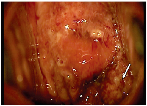
Fig. 1 Colposcopic finding of uterine cervix. Note a mass 2.5 cm in maximal dimension that involved the left posterior fornix (arrow).
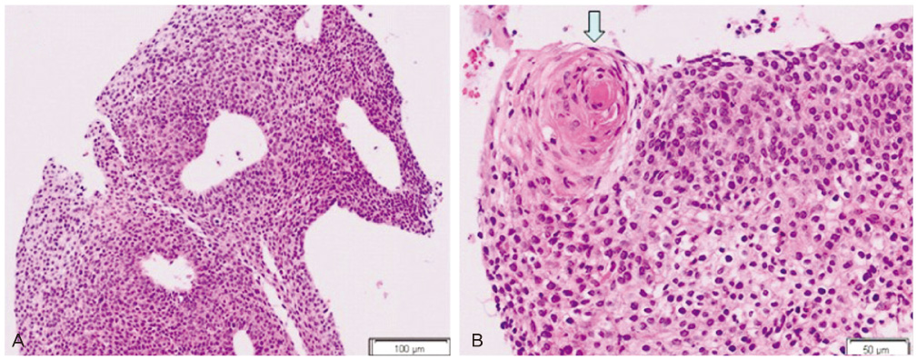
Fig. 2 (A) Transitional cells with papillary figures are seen (H&E, ×100). (B) Focally keratin forming squamous cell nest is seen (H&E, ×200).

Fig. 3 (A) Immunohistochemistry for cytokeratin (CK) 20 is negative (peroxidase, DAB; ×200). (B) Immunohistochemistry for CK 7 is positive in the cytoplasm and cytoplasmic membrane (peroxidase, DAB; ×200).
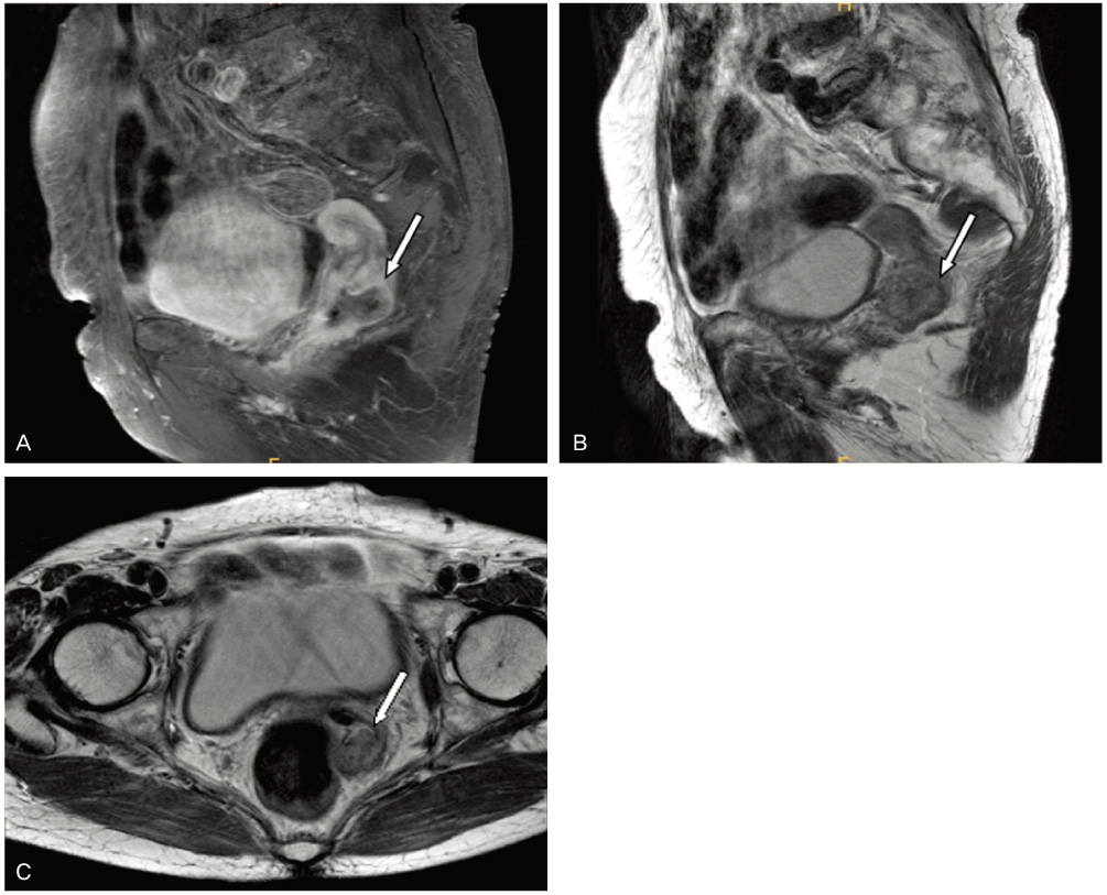
Fig. 4 Magnetic resonance imaging shows ill-defined mass lesion in uterine cervix posterior aspect. Which lesion measured about 2.3 × 2.0 × 1.7 cm (arrow) (A, T2WI sagittal scanning; B, T1WI sagittal scanning; C, T1WI axial scanning).
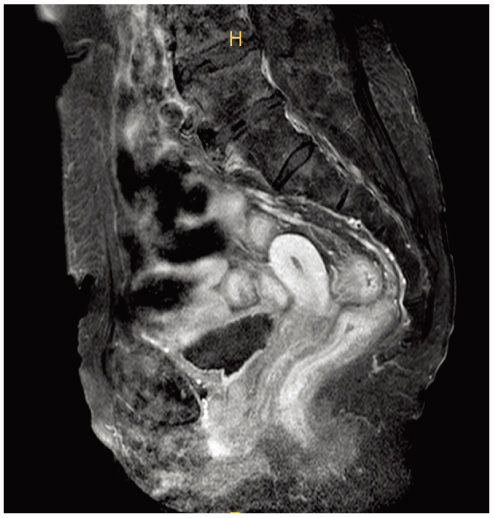
Fig. 5 Magnetic resonance imaging shows no definite mass lesion delineated in uterine cervix (T2WI sagittal scanning).
Reference
-
1. Rose PG, Stoler MH, Abdul-Karim FW. Papillary squamotransitional cell carcinoma of the vagina. Int J Gynecol Pathol. 1998. 17:372–375.2. Vesoulis Z, Erhardt CA. Cytologic diagnosis of vaginal papillary squamotransitional cell carcinoma. A case report. Acta Cytol. 2001. 45:465–469.3. Gao Z, Bhuiya T, Falkowski O. Papillary squamotransitional cell carcinoma of the vagina: a case report and review of literature. J Obstet Gynaecol. 2005. 25:94–96.4. Albores-Saavedra J, Young RH. Transitional cell neoplasms (carcinomas and inverted papillomas) of the uterine cervix. A report of five cases. Am J Surg Pathol. 1995. 19:1138–1145.5. Randall ME, Andersen WA, Mills SE, Kim JA. Papillary squamous cell carcinoma of the uterine cervix: a clinicopathologic study of nine cases. Int J Gynecol Pathol. 1986. 5:1–10.6. Koenig C, Turnicky RP, Kankam CF, Tavassoli FA. Papillary squamotransitional cell carcinoma of the cervix: a report of 32 cases. Am J Surg Pathol. 1997. 21:915–921.7. Fetissof F, Haillot O, Lanson Y, Arbeille B, Lansac J. Papillary tumour of the vagina resembling transitional cell carcinoma. Pathol Res Pract. 1990. 186:358–364.8. Jendresen MB, Kvist E, Glenthøj A. Papillary transitional cell tumour in the vagina. Scand J Urol Nephrol. 1997. 31:107–108.9. Soslow RA, Rouse RV, Hendrickson MR, Silva EG, Longacre TA. Transitional cell neoplasms of the ovary and urinary bladder: a comparative immunohistochemical analysis. Int J Gynecol Pathol. 1996. 15:257–265.10. Ramaekers F, Huysmans A, Schaart G, Moesker O, Vooijs P. Tissue distribution of keratin 7 as monitored by a monoclonal antibody. Exp Cell Res. 1987. 170:235–249.11. Schaafsma HE, Ramaekers FC, van Muijen GN, Lane EB, Leigh IM, Robben H, et al. Distribution of cytokeratin polypeptides in human transitional cell carcinomas, with special emphasis on changing expression patterns during tumor progression. Am J Pathol. 1990. 136:329–343.12. Moll R, Löwe A, Laufer J, Franke WW. Cytokeratin 20 in human carcinomas. A new histodiagnostic marker detected by monoclonal antibodies. Am J Pathol. 1992. 140:427–447.13. Lininger RA, Ashfaq R, Albores-Saavedra J, Tavassoli FA. Transitional cell carcinoma of the endometrium and endometrial carcinoma with transitional cell differentiation. Cancer. 1997. 79:1933–1943.14. Singer G, Hohl MK, Hering F, Anabitarte M. Transitional cell carcinoma of the vagina with pagetoid spread pattern. Hum Pathol. 1998. 29:299–301.15. Ollayos CW, Lichy J, Duncan BW, Ali IS. Papillary squamous cell carcinoma of the uterine cervix: report of a case with HPV 16 DNA and brief review. Gynecol Oncol. 1996. 63:388–391.
- Full Text Links
-
- Actions
-
Cited
- CITED
-
- Close
- Share
-
- Similar articles
-
- A Case of Papillary Type of Renal Cell Carcinoma after Renal Injury in a Child
- A case of primary squamous cell carcinoma of vagina
- Papillary Transitional Cell Carcinoma of the Bladder: Report of a Case
- Concurrent Medullay and Papillary Carcinoma of the Thyroid
- Fine Needle Aspiration Cytology of Papillary Renal Cell Carcinoma: A Case Report
