Korean Circ J.
2013 Oct;43(10):702-704. 10.4070/kcj.2013.43.10.702.
Isolated, Broad-Based Apical Diverticulum: Cardiac Magnetic Resonance Is a "Terminator" of Cardiac Imaging Modality for the Evaluation of Cardiac Apex
- Affiliations
-
- 1Cardiovascular Center, Seoul National University Hospital, Seoul, Korea. cardiman73@gmail.com
- 2Department of Internal Medicine, Seoul National University College of Medicine, Seoul, Korea.
- 3Department of Radiology, Seoul National University College of Medicine, Seoul, Korea.
- KMID: 1826563
- DOI: http://doi.org/10.4070/kcj.2013.43.10.702
Abstract
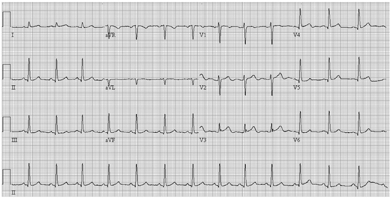
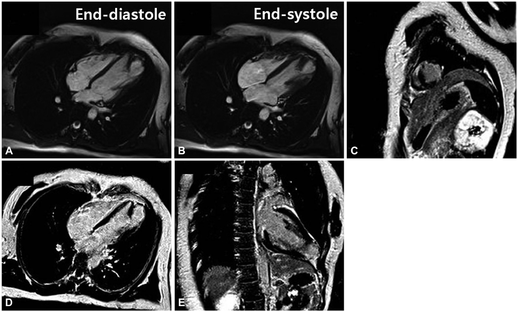
- In spite of the frequent involvement of many cardiac diseases, it is difficult to evaluate the left ventricular apex in detail with transthoracic echocardiography, a first-line imaging modality in cardiovascular diseases, because the apex is very closely located at the echocardiographic probe. Cardiac magnetic resonance enables us to evaluate the cardiac apex without any limitation to the image acquisition. We here present a case regarding a broad-based apical diverticulum, which was initially confused with apical aneurysm.
MeSH Terms
Figure
Reference
-
1. O'Bryan . Peacock TB, editor. On Malformations of the Human Heart. 2nd ed. London, UK: Churchill & Sons;1866.2. Paz Y, Fridman E, Shakalia FM, Danieli J, Mishaly D. Repair of an isolated huge congenital left ventricular diverticulum. J Thorac Cardiovasc Surg. 2004; 128:313–314.3. Ohlow MA. Congenital left ventricular aneurysms and diverticula: definition, pathophysiology, clinical relevance and treatment. Cardiology. 2006; 106:63–72.4. Weinsaft JW, Kim HW, Crowley AL, et al. LV thrombus detection by routine echocardiography: insights into performance characteristics using delayed enhancement CMR. JACC Cardiovasc Imaging. 2011; 4:702–712.5. Kim RJ, Fieno DS, Parrish TB, et al. Relationship of MRI delayed contrast enhancement to irreversible injury, infarct age, and contractile function. Circulation. 1999; 100:1992–2002.6. Moon JC, Reed E, Sheppard MN, et al. The histologic basis of late gadolinium enhancement cardiovascular magnetic resonance in hypertrophic cardiomyopathy. J Am Coll Cardiol. 2004; 43:2260–2264.
- Full Text Links
- Actions
-
Cited
- CITED
-
- Close
- Share
- Similar articles
-
- Isolated Congenital Left Ventricular Diverticulum in Adults
- High field strength magnetic resonance imaging of cardiovascular diseases
- A Case of Isolated Congenital Right Ventricular Diverticulum in Adult
- Typical and Atypical Magnetic Resonance Imaging Appearances of Primary Cardiac Lymphoma
- Using CT to Evaluate Cardiac Function


