A Case of Three Consecutive Events of Acute Myocardial Infarctions in Three Different Vessels
- Affiliations
-
- 1Division of Cardiology, Department of Internal Medicine, Daejeon St. Mary's Hospital, College of Medicine, The Catholic University of Korea, Daejeon, Korea. hhhsungho@naver.com
- 2Division of Thoracic and Cardiovascular Surgery, Daejeon St. Mary's Hospital, College of Medicine, The Catholic University of Korea, Daejeon, Korea.
- KMID: 1826561
- DOI: http://doi.org/10.4070/kcj.2013.43.10.694
Abstract
- A 51-year-old man was being admitted to the emergency department with chest pains. He had a history of acute myocardial infarction (MI) on two prior occasions and was successfully treated with drug eluting stents. He was diagnosed with 3 consecutive events of acute MI in 3 different vessels. The consecutive events of acute MI in different vessels are a very rare case. He did not have risk factors, such as coagulation abnormality, clopidogrel resistance, patient's compliance and vessel abnormality, except for his cigarette smoking. We reported the first case with 3 consecutive events of acute MI in each 3 vessels during a long-term interval.
Keyword
MeSH Terms
Figure
-
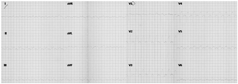
Fig. 1 ECG on February 21, 2000. The ECG shows ST-segment elevation in the inferior leads (II and aVF) and the anterior (V 2-4) leads. ECG: electrocardiogram.
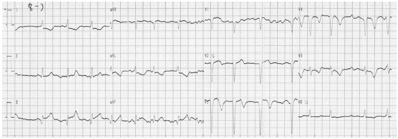
Fig. 2 ECG on December 30, 2007. The ECG shows ST-segment elevation in the inferior (II, III, and aVF) leads. ECG: electrocardiogram.

Fig. 3-1 Right coronary angiography on December 30, 2007. A: the initial coronary angiography shows a total occlusion with thrombus and TIMI 0 distal flow in the mid portion of the RCA (white arrow). B: the post stent coronary angiography shows TIMI 3 distal flow in the RCA, which is successfully treated with a paclitaxel eluting stent (white arrow). TIMI: Thrombolysis in Myocardial Infarction, RCA: right coronary artery.

Fig. 3-2 Coronary angiography on December 30, 2007. A right anterior oblique view. A: the initial coronary angiography shows a 95% restenosis in stent with thrombus and TIMI 0 distal flow in the proximal portion of the LAD (white arrow). B: the post stent coronary angiography shows TIMI 3 distal flow in the LAD, which is successfully treated with a zotarolimus eluting stent (black arrow) and overlapping a sirolimus eluting stent (white arrow). TIMI: Thrombolysis in Myocardial Infarction, LAD: left anterior descending coronary artery.

Fig. 4 ECG on September 25, 2012. The ECG shows Q-wave and ST elevation in V 1-5 and T-inversion in V 4-6. ECG: electrocardiogram.
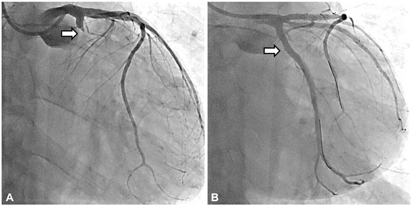
Fig. 5 Coronary angiography on September 25, 2012. A left anterior oblique view. A: the initial coronary angiography shows a total occlusion with thrombus and TIMI 0 distal flow in the proximal portion of the LCX (white arrow). B: the post stent coronary angiography shows TIMI 3 distal flow in the LCX, which is successfully treated with a zotarolimus eluting stent (white arrow). TIMI: Thrombolysis in Myocardial Infarction, LCX: left circumflex coronary artery.
Reference
-
1. Godino C, Mendolicchio L, Figini F, et al. Comparison of VerifyNow-P2Y12 test and Flow Cytometry for monitoring individual platelet response to clopidogrel. What is the cut-off value for identifying patients who are low responders to clopidogrel therapy? Thromb J. 2009; 7:4.2. Virmani R, Kolodgie FD, Burke AP, Farb A, Schwartz SM. Lessons from sudden coronary death: a comprehensive morphological classification scheme for atherosclerotic lesions. Arterioscler Thromb Vasc Biol. 2000; 20:1262–1275.3. Burke AP, Farb A, Malcom GT, Liang Y, Smialek JE, Virmani R. Plaque rupture and sudden death related to exertion in men with coronary artery disease. JAMA. 1999; 281:921–926.4. Holmes DR Jr, Kereiakes DJ, Garg S, et al. Stent thrombosis. J Am Coll Cardiol. 2010; 56:1357–1365.5. Wang TH, Bhatt DL, Topol EJ. Aspirin and clopidogrel resistance: an emerging clinical entity. Eur Heart J. 2006; 27:647–654.6. Michos ED, Ardehali R, Blumenthal RS, Lange RA, Ardehali H. Aspirin and clopidogrel resistance. Mayo Clin Proc. 2006; 81:518–526.7. Sweeny JM, Gorog DA, Fuster V. Antiplatelet drug 'resistance'. Part 1: mechanisms and clinical measurements. Nat Rev Cardiol. 2009; 6:273–282.8. Kanei Y, Janardhanan R, Fox JT, Gowda RM. Multivessel coronary artery thrombosis. J Invasive Cardiol. 2009; 21:66–68.9. Falk E. Multiple culprits in acute coronary syndromes: systemic disease calling for systemic treatment. Ital Heart J. 2000; 1:835–838.10. Gan F, Hu D, Dai T. Acute multivessel coronary artery occlusion: a case report. BMC Res Notes. 2012; 5:523.11. Kim SS, Jeong MH, Kim HK, et al. Acute and subacute stent thrombosis in a patient with clopidogrel resistance: a case report. Korean Circ J. 2009; 39:434–438.12. Brito JD, Almeida MS, Ribeiras R, Melo JQ, Almeida RH, Silva JA. Recurrent myocardial infarction in a patient with papillary fibroelastoma. Arq Bras Cardiol. 2012; 98:e7–e10.13. Peovska I, Maksimovic J, Kalpak O, Pejkov H, Bosevski M. Recurrent myocardial infarction in a young football player with antithrombin III deficiency. Cardiol J. 2008; 15:463–466.14. Chang MH, Bang SY, Kim TH, et al. Recurrent acute myocardial infarctions and Budd-Chiari syndrome in young woman with Behcet's disease. J Korean Rheum Assoc. 2007; 14:96–100.
- Full Text Links
-
- Actions
-
Cited
- CITED
-
- Close
- Share
-
- Similar articles
-
- Acute Myocardial Infarction with Microthrombi in Cardiac Small Vessels after COVID-19 Vaccination (ChAdOx1 nCov-19): A Case Report
- Drug-Eluting Stent Used to Treat a Case of Recurrent Right Coronary Artery In-Stent Restenoses often Accompanied by Acute Inferior Wall Myocardial Infarction
- Early Result of Surgical Revascularization for Acute Myocardial Infarction
- The Prognostic Significance of Maximal Precordial ST-Segment Depression in Patients with Acute Inferior Myocardial Infarction
- Two Cases of Acute Myocardial Infarctions with Normal Coronary Arteriograms and Early Complete Recovery of Myocardial Dysfunction
