Spinal Capillary Hemangioma Involving the Lumbar Epidural and Paraspinal Spaces: A Case Report
- Affiliations
-
- 1Department of Radiology, Hanyang University Medical Center, Hanyang University College of Medicine, Seoul, Korea. yjleeee@hanyang.ac.kr
- 2Department of Radiology, Hanyang University Guri Hospital, Hanyang University College of Medicine, Seoul, Korea.
- 3Department of Pathology, Hanyang University Medical Center, Hanyang University College of Medicine, Seoul, Korea.
- KMID: 1823920
- DOI: http://doi.org/10.3348/jksr.2015.73.1.36
Abstract
- Spinal capillary hemangiomas in the epidural space are extremely rare; however, a preoperative radiological diagnosis is very important because of the risk of massive intraoperative hemorrhage. We report a case of a spinal capillary hemangioma involving the lumbar epidural and paraspinal spaces.
Figure
-
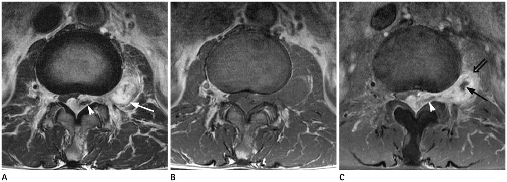
Fig. 1 Lumbar MRI of a 54-year-old man presenting lower extremity weakness. A Dumbbell-shaped mass involving both epidural and paraspinal space is noted. On T2-weighted axial image (A), hemorrhagic and cystic portion of the mass are shown as a low signal intensity area (arrowhead) and a high signal intensity area (arrow), respectively. On pre-contrast axial T1-weighted image (B), the mass is generally iso-intense to adjacent muscle with some heterogeneity. On post-contrast axial T1-weighted image (C), the mass enhances well with areas of non-enhancement suggesting hemorrhagic (arrowhead) and cystic portion (arrow). At anterior aspect of paraspinal mass, partially infiltrative portion is noted (double arrow).
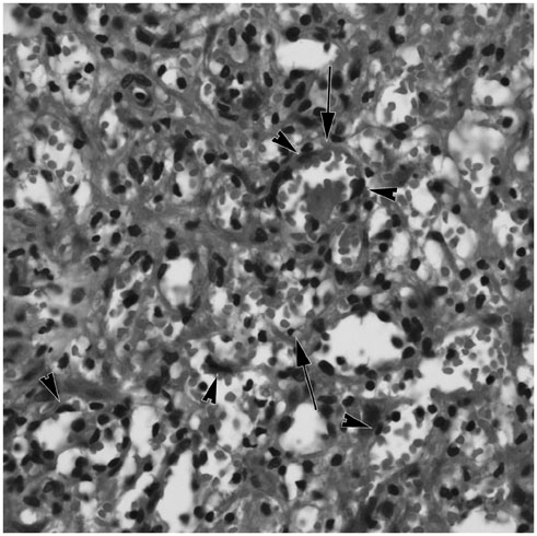
Fig. 2 A photograph show numerous, tightly packed, capillary size vessels (arrows) that were lined by a single layer of cytologically benign endothelial cells (arrowheads) (H&E, × 400 section).
Reference
-
1. Choi BY, Chang KH, Choe G, Han MH, Park SW, Yu IK, et al. Spinal intradural extramedullary capillary hemangioma: MR imaging findings. AJNR Am J Neuroradiol. 2001; 22:799–802.2. Kasukurthi R, Ray WZ, Blackburn SL, Lusis EA, Santiago P. Intramedullary capillary hemangioma of the thoracic spine: case report and review of the literature. Rare Tumors. 2009; 1:e10.3. Gencpinar P, Açıkbas¸ SC, Nur BG, Karaali K, Arslan M, Gurer EI, et al. Epidural capillary hemangioma: a review of the literature. Clin Neurol Neurosurg. 2014; 126:99–102.4. Nowak DA, Widenka DC. Spinal intradural capillary haemangioma: a review. Eur Spine J. 2001; 10:464–472.5. Tekin T, Bayrakli F, Simsek H, Colak A, Kutlay M, Demircan MN. Lumbar epidural capillary hemangioma presenting as lumbar disc herniation disease: case report. Spine (Phila Pa 1976). 2008; 33:E795–E797.6. Shin JH, Lee HK, Jeon SR, Park SH. Spinal intradural capillary hemangioma: MR findings. AJNR Am J Neuroradiol. 2000; 21:954–956.7. Caruso G, Galarza M, Borghesi I, Pozzati E, Vitale M. Acute presentation of spinal epidural cavernous angiomas: case report. Neurosurgery. 2007; 60:E575–E576. discussion E5768. Jo BJ, Lee SH, Chung SE, Paeng SS, Kim HS, Yoon SW, et al. Pure epidural cavernous hemangioma of the cervical spine that presented with an acute sensory deficit caused by hemorrhage. Yonsei Med J. 2006; 47:877–880.9. Calonje E, Mentzel T, Fletcher CD. Pseudomalignant perineurial invasion in cellular ('infantile') capillary haemangiomas. Histopathology. 1995; 26:159–164.10. Kivrak AS, Koc O, Emlik D, Kiresi D, Odev K, Kalkan E. Differential diagnosis of dumbbell lesions associated with spinal neural foraminal widening: imaging features. Eur J Radiol. 2009; 71:29–41.
- Full Text Links
-
- Actions
-
Cited
- CITED
-
- Close
- Share
-
- Similar articles
-
- Epidural Hemangioma: A Case Report
- Two Cases of Epidural Cavernous Hemangioma in the Thoraic Spine
- Spinal Epidural Cavernous Hemangioma of the Upper Thoracic Vertebrae: Case Report
- Pure Extradural Cavernous Hemangioma Presenting as a Lumbar Radiculopathy
- Lumbar Paraspinal Myonecrosis Following Combined Spinal Epidural Anesthesia: A case report
