J Korean Soc Spine Surg.
2009 Dec;16(4):294-298. 10.4184/jkss.2009.16.4.294.
Surgical Treatment of Ossifying Fibromyxoid Tumor Invading the Lumbar Spine: A Case Report
- Affiliations
-
- 1Department of Orthopedic Surgery, Samsung Medical Center, Sungkyunkwan University, School of Medicine, Seoul, Korea.
- 2Department of Orthopedic Surgery, Wonkwang University Sanbon Hospital, Gunpo, Korea. niceo@daum.net
- KMID: 1819812
- DOI: http://doi.org/10.4184/jkss.2009.16.4.294
Abstract
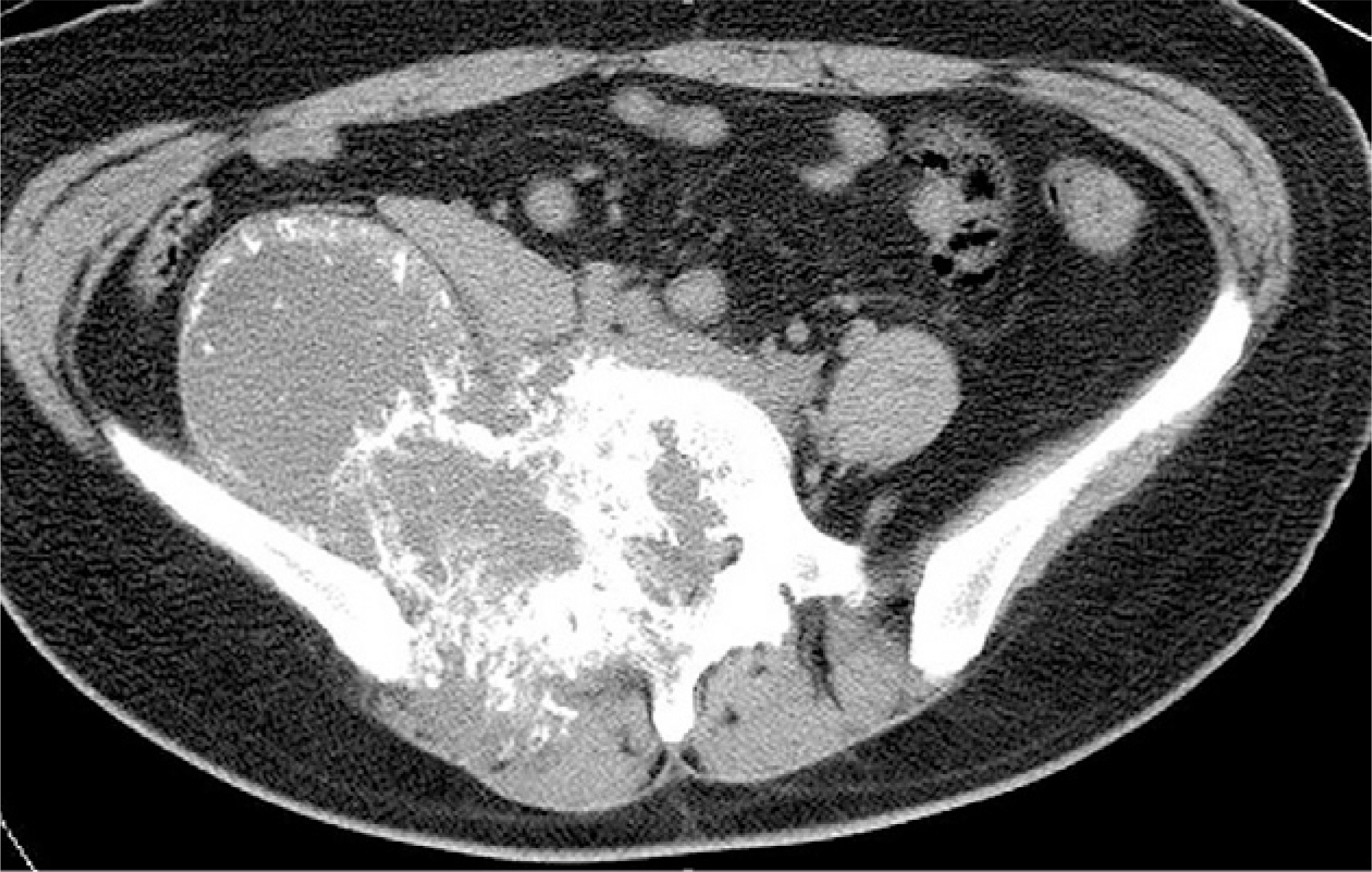
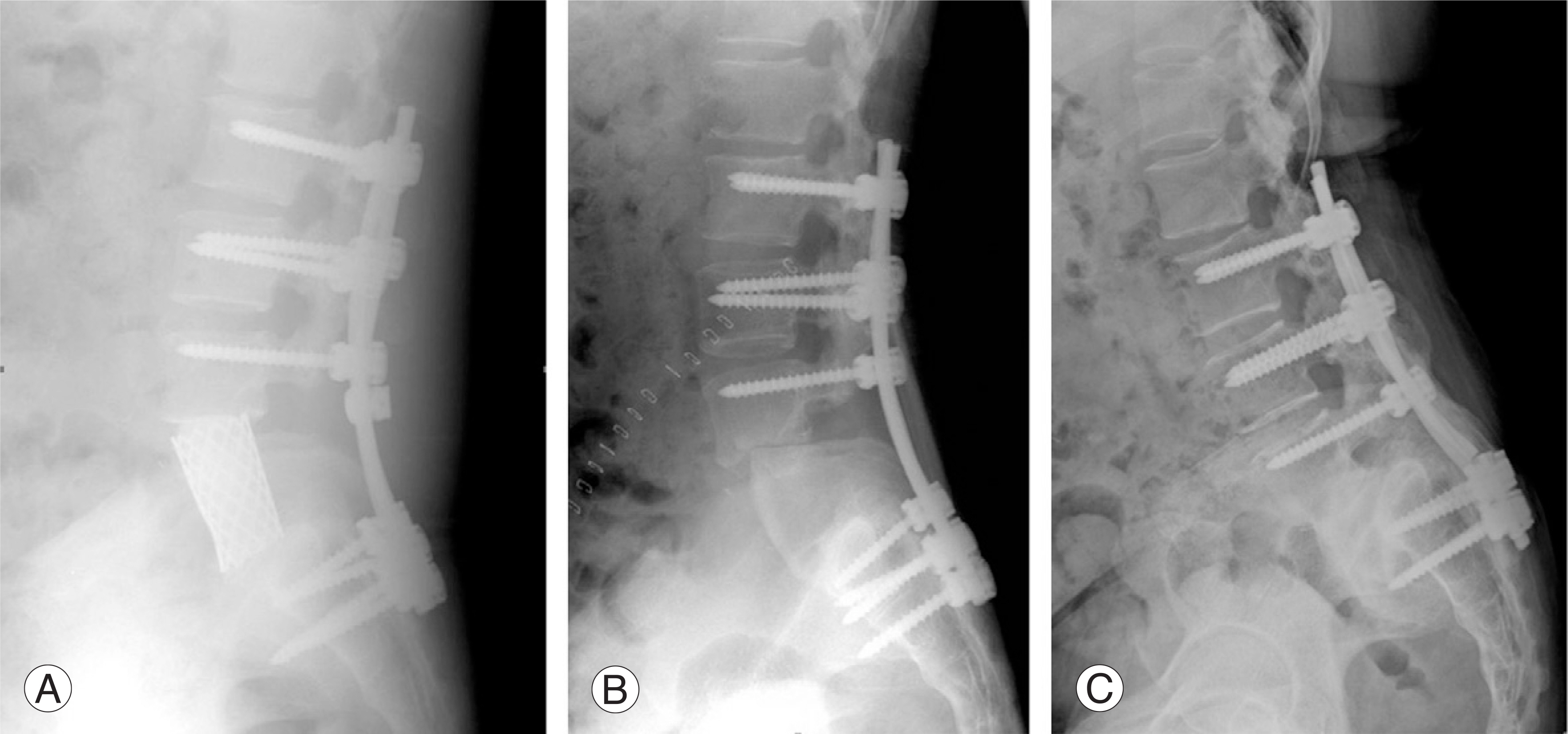
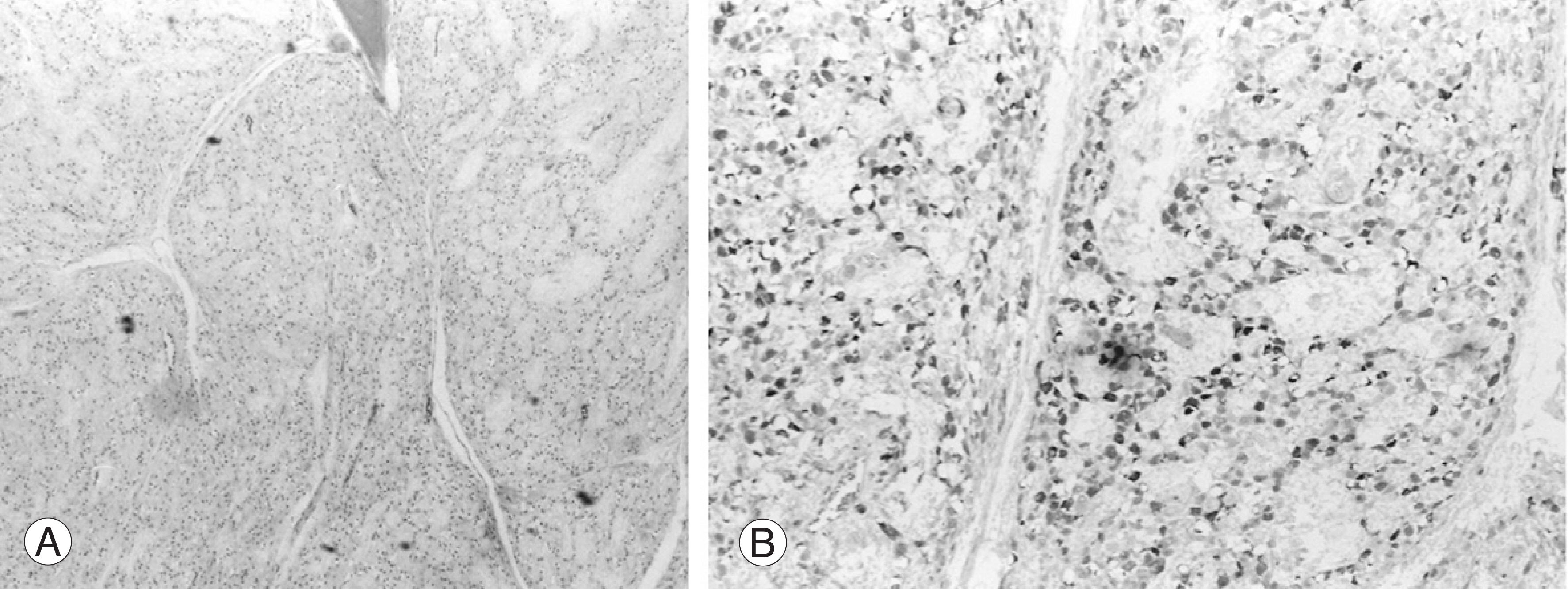
- Ossifying fibromyxoid tumor is rare soft tissue neoplasm of an uncertain histogenesis, and this was first described in 1989. The majority of the reported cases have involved the soft tissue of the extremities. We present here on a case of atypical ossifying fibromixoid tumor that had invaded the spine and we report on its management and outcome. We also review the relevant literature.
MeSH Terms
Figure
Reference
-
01). Enzinger FM., Weiss SW., Liang CY. Ossifying fibromyxoid tumor of soft parts. A clinicopathological analysis of 59 cases. Am J Surg Pathol. 1989. 13:817–827.02). Schaffler G,Raith J., Ranner G., Weybora W., Jeserschek R. Radiographic appearance of an ossifying fibromyxoid tumor of soft parts. Skeletal Radiol. 1997. 26:615–618.03). Ogose A., Otsuka H., Morita T., Kobayashi H., Hirata Y. Ossifying fibromyxoid tumor resembling parosteal osteosarcoma. Skeletal Radiol. 1998. 27:578–580.
Article04). Folpe AL., Weiss SW. Ossifying fibromyxoid tumor of soft parts: a clinicopathologic study of 70 cases with emphasis on atypical and malignant variants. Am J Surg Pathol. 2003. 27:421–431.05). Miettinen M., Finnell V., Fetsch JF. Ossifying fibromyxoid tumor of soft parts—a clinicopathologic and immunohistochemical study of 104 cases with long-term follow-up and a critical review of the literature. Am J Surg Pathol. 2008. 32:996–1005.
Article06). Donner LR. Ossifying fibromyxoid tumor of soft parts: evidence supporting Schwann cell origin. Hum Pathol. 1992. 23:200–202.
Article07). Schofield JB., Krausz T., Stamp GW., Fletcher CD., Fisher C., Azzopardi JG. Ossifying fibromyxoid tumour of soft parts: immunohistochemical and ultrastructural analysis. Histopathology. 1993. 22:101–112.
Article08). Sovani V., Velagaleti GV., Filipowicz E., Gatalica Z., Knisely AS. Ossifying fibromyxoid tumor of soft parts: report of a case with novel cytogenetic findings. Cancer Genet Cytogenet. 2001. 127:1–6.09). Nishio J., Iwasaki H., Ohjimi Y, et al. Ossifying fibromyx-oid tumor of soft parts. Cytogenetic findings. Cancer Genet Cytogenet. 2002. 133:124–128.10). Oqilvie CM., Torbert JT., Finstein JL, et al. Clinical utility of percutaneous biopsies of musculoskeletal tumors. Clin Orthop Relat Res. 2006. 450:95–100.


