J Korean Soc Radiol.
2014 Aug;71(2):80-83. 10.3348/jksr.2014.71.2.80.
Bilateral Ureteral Calcifications Associated with Systemic Lupus Erythematosus: A Case Report
- Affiliations
-
- 1Department of Radiology, Yeungnam University Hospital, Daegu, Korea.
- 2Department of Radiology, Dongsan Medical Center, Keimyung University College of Medicine, Daegu, Korea. kseehdr@dsmc.or.kr
- 3Department of Radiology, Kyungpook National University Hospital, Daegu, Korea.
- KMID: 1819784
- DOI: http://doi.org/10.3348/jksr.2014.71.2.80
Abstract
- A 23-year-old woman with systematic lupus erythematosus (SLE) presented with left flank pain and hematuria, after seven months of hospital treatment with prednisolone. Computed tomography (CT) of abdomen showed multifocal calcifications of bilateral ureteral walls and hydronephrosis by left ureteral stenosis. These calcifications may be associated with lupus-induced inflammatory reaction of ureteral wall. In this study, we also present review of the literature associated with the similar cases.
MeSH Terms
Figure
-
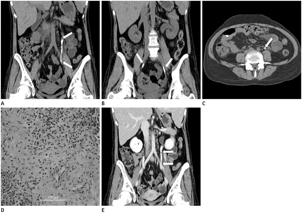
Fig. 1 A 23-year-old female patient with systematic lupus erythematosus. A-C. Non-contrast coronal CT scans (A, B) show multifocal calcifications (arrows) in both ureters. Non-contrast axial CT scan (C) shows calcifications with central lucency (arrow) in left ureter, consistent with calcifications of ureteral wall. D. Microscopy shows edema and mixed inflammatory cells infiltration of the lamina propria mainly consisting of lymphocytes (H&E, × 100 original magnification). The calcifications are also noted at lamina propria of ureteral wall (not shown). E. Follow-up coronal CT scan after 6 months later shows slight aggravation of multifocal calcifications of left ureteral wall (arrows).
Reference
-
1. Jiang DX, Liao Y, Bai YJ. Calcification of bilateral ureters: a novel association with systemic lupus erythematosus. Chin Med J (Engl). 2012; 125:2235–2223.2. Tristano AG, Villarroel JL, Rodríguez MA, Millan A. Calcinosis cutis universalis in a patient with systemic lupus erythematosus. Clin Rheumatol. 2006; 25:70–74.3. Mok CC, Lau CS. Pathogenesis of systemic lupus erythematosus. J Clin Pathol. 2003; 56:481–490.4. Asia S, Martellotto G, Belén R, Sesín AM, Gamróm S, Drenkard C. Obstructive uropathy as the only manifestation of flare in a patient with systemic lupus erythematosus and anti-phospholipid syndrome. Lupus. 2008; 17:46–49.5. Wang LJ, Wu CF, Wong YC, Chuang CK, Chu SH, Chen CJ. Imaging findings of urinary tuberculosis on excretory urography and computerized tomography. J Urol. 2003; 169:524–528.6. Shebel HM, Elsayes KM, Abou El Atta HM, Elguindy YM, El-Diasty TA. Genitourinary schistosomiasis: life cycle and radiologic-pathologic findings. Radiographics. 2012; 32:1031–1046.7. Thomas SD, Sanders PW 3rd, Pollack H. Primary amyloidosis of urinary bladder and ureter: cause of mural calcification. Urology. 1977; 9:586–589.8. Gold RP, Saitas V, Pellman C. Congenital ureteropelvic junction obstruction with calcified renal pelvis and superimposed spindle cell urothelial carcinoma. Urol Radiol. 1990; 12:15–17.9. Masuda Y, Uchiyama Y, Hashimoto S, Uchiyama S, Iwata M. Symmetrical progressive intracranial calcification in a patient with SLE. Intern Med. 2010; 49:351.10. Okada J, Nomura M, Shirataka M, Kondo H. Prevalence of soft tissue calcifications in patients with SLE and effects of alfacarcidol. Lupus. 1999; 8:456–461.
- Full Text Links
-
- Actions
-
Cited
- CITED
-
- Close
- Share
-
- Similar articles
-
- Bilateral ureteral calcification with left kidney hydronephrosis in systemic lupus erythematosus
- A Case of Transverse Myelitis as a First Manifestation of Systemic Lupus Erythematosus
- Multiple Dermatofibromas in a woman with Systemic Lupus Erythematosus
- A Case Of Systemic Lupus Erythematosus Associated With Hyperthyroidism And Severe Retinopathy
- A Case of Lupus Enteritis That Developed during the Treatment of Systemic Lupus Erythematosus
