J Korean Neurosurg Soc.
2013 Aug;54(2):139-141. 10.3340/jkns.2013.54.2.139.
Hypertensive Encephalopathy with Reversible Brainstem Edema
- Affiliations
-
- 1Department of Neurosurgery, Seoul National University College of Medicine, Seoul, Korea.
- 2Department of Neurosurgery, The Armed Forces Capital Hospital, Seongnam, Korea.
- 3Department of Neurosurgery, The Catholic University of Korea, Bucheon St. Mary's Hospital, Bucheon, Korea. armada1997@naver.com
- KMID: 1814253
- DOI: http://doi.org/10.3340/jkns.2013.54.2.139
Abstract
- Presented here is a 36-year-old male with arterial hypertension who developed brainstem edema and intracranial hemorrhage. Magnetic resonance scan revealed diffuse brainstem hyperintensity in T2-weighted and fluid-attenuated inversion-recovery images, with an increase in apparent diffusion coefficient values. After a reduction in blood pressure, rapid resolution of the brainstem edema was observed on follow-up. The patient's condition was thus interpreted as hypertensive brainstem encephalopathy. While many consider this a vasogenic phenomenon, induced by sudden, severe hypertension, the precise mechanism remains unclear. Prompt recognition and aggressive antihypertensive treatment in such patients are essential to prevent permanent or life-threatening neurologic injury.
Keyword
MeSH Terms
Figure
-
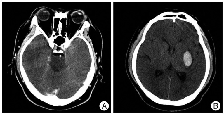
Fig. 1 Axial CT image of brain showing intracranial hemorrhage of left basal ganglia with brainstem hypodensity (A and B).

Fig. 2 Axial MRI T2 and fluid-attenuated inversion recovery image showing increased signal intensity of pons and midbrain (A and B) : increase in apparent diffusion coefficient values for same area (C) and there are no abnormal findings in subcortical white matter of the parietal and occipital lobes other than the small amount of basal ganglia (D). MRI and diffusion-weighted images after blood pressure stabilization; note resolution of prior brainstem abnormalities (E and F).
Reference
-
1. Bartynski WS. Posterior reversible encephalopathy syndrome, part 2 : controversies surrounding pathophysiology of vasogenic edema. AJNR Am J Neuroradiol. 2008; 29:1043–1049. PMID: 18403560.
Article2. Chang GY, Keane JR. Hypertensive brainstem encephalopathy : three cases presenting with severe brainstem edema. Neurology. 1999; 53:652–654. PMID: 10449143.
Article3. Chen TY, Lee HJ, Wu TC, Tsui YK. MR imaging findings of medulla oblongata involvement in posterior reversible encephalopathy syndrome secondary to hypertension. AJNR Am J Neuroradiol. 2009; 30:755–757. PMID: 18854436.
Article4. Doi Y, Kimura F, Fujiyama T, Fujimura C, Nishina T, Sato T, et al. Hypertensive brainstem encephalopathy without parieto-occipital lesion--two case reports. Neurol Med Chir (Tokyo). 2006; 46:75–79. PMID: 16498216.
Article5. Garg RK. Posterior leukoencephalopathy syndrome. Postgrad Med J. 2001; 77:24–28. PMID: 11123390.
Article6. Hinchey J, Chaves C, Appignani B, Breen J, Pao L, Wang A, et al. A reversible posterior leukoencephalopathy syndrome. N Engl J Med. 1996; 334:494–500. PMID: 8559202.
Article7. Ijima T, Kubota Y, Kuroiwa T, Sankawa H. Blood-brain barrier opening following transient reflex sympathetic hypertension. Acta Neurochir Suppl (Wien). 1994; 60:142–144. PMID: 7976528.
Article8. Legriel S, Pico F, Azoulay E. Understanding Posterior Reversible Encephalopathy Syndrome. Ann Upd Intens Care Emerg Med. 2011; 1:631–653.
Article
- Full Text Links
-
- Actions
-
Cited
- CITED
-
- Close
- Share
-
- Similar articles
-
- Two Cases of Hypertensive Encephalopathy Involving the Brainstem
- Hypertensive Brainstem Encephalopathy Combined with Acute Ischemic Stroke
- Reversible Obstructive Hydrocephalus Associated with Hypertensive Brainstem Encephalopathy
- Predominant Hypertensive Brainstem Encephalopathy with Supratentorial Involvement: Case Report and Literature Review
- Reversible Pontine MRI Lesion in Acute Thalamic Infarct: Reversible Encephalopathy due to Hypertension?
