The Usefulness of Brain Magnetic Resonance Imaging with Mild Head Injury and the Negative Findings of Brain Computed Tomography
- Affiliations
-
- 1Department of Neurosurgery, Seoul Medical Center, Seoul, Korea. davidmhkong@gmail.com
- KMID: 1814245
- DOI: http://doi.org/10.3340/jkns.2013.54.2.100
Abstract
OBJECTIVE
To investigate the cases of intracranial abnormal brain MRI findings even in the negative brain CT scan after mild head injury.
METHODS
During a 2-year period (January 2009-December 2010), we prospectively evaluated both brain CT and brain MRI of 180 patients with mild head injury. Patients were classified into two groups according to presence or absence of abnormal brain MRI finding even in the negative brain CT scan after mild head injury. Two neurosurgeons and one neuroradiologist validated the images from both brain CT scan and brain MRI double blindly.
RESULTS
Intracranial injury with negative brain CT scan after mild head injury occurred in 18 patients (10.0%). Headache (51.7%) without neurologic signs was the most common symptom. Locations of intracranial lesions showing abnormal brain MRI were as follows; temporal base (n=8), frontal pole (n=5), falx cerebri (n=2), basal ganglia (n=1), tentorium (n=1), and sylvian fissure (n=1). Intracranial injury was common in patients with a loss of consciousness, symptom duration >2 weeks, or in cases of patients with linear skull fracture (p=0.00013), and also more frequent in multiple associated injury than simple one (35.7%>8.6%) (p=0.105).
CONCLUSION
Our investigation showed that patients with mild head injury even in the negative brain CT scan had a few cases of intracranial injury. These findings indicate that even though the brain CT does not show abnormal findings, they should be thoroughly watched in further study including brain MRI in cases of multiple injuries and when their complaints are sustained.
MeSH Terms
Figure
-
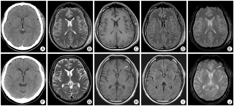
Fig. 1 Illustration by magnetic resonance imaging (MRI) compared to computed tomography (CT) in 2 study participants of mild head injury. A 22-year-old male corresponded brain CT (-) and brain MRI (-) group I (A-E) which had no brain CT or MRI parenchymal lesion except extensive scalp swelling (*) of left parieto-occipital area and subgaleal hematoma after mild head injury. A 46-year-old male demonstrated initial brain CT (F) was normal after traffic accident and MRI at post-injury 7 days showed minimal subdural hematoma (arrow) on left temporo-parietal lobe at axial section of T1-weighted image (H) and the hemorrhagic contusion on left frontal lobe (arrow) at axial T2-weighted gradient echo MRI image (J) which corresponded brain CT (-) and brain MRI (+) group II (F-J).
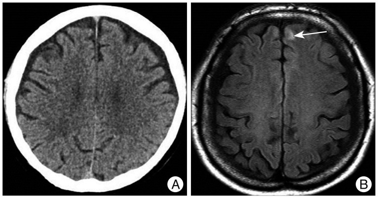
Fig. 2 Computed tomography scan showing not abnormal intracranial pathology (A) and T1-weighted MR image showing hemorrhagic contusion (arrow) on left frontal area (B) which was classified as group II.
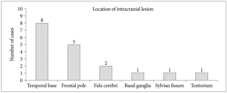
Fig. 3 The numbers of the location of intracranial lesions of the group II.
Reference
-
1. Alahmadi H, Vachhrajani S, Cusimano MD. The natural history of brain contusion : an analysis of radiological and clinical progression. J Neurosurg. 2010; 112:1139–1145. PMID: 19575576.
Article2. Bordignon KC, Arruda WO. CT scan findings in mild head trauma : a series of 2,000 patients. Arq Neuropsiquiatr. 2002; 60:204–210. PMID: 12068346.3. Borg J, Holm L, Cassidy JD, Peloso PM, Carroll LJ, von Holst H, et al. Diagnostic procedures in mild traumatic brain injury : results of the WHO Collaborating Centre Task Force on Mild Traumatic Brain Injury. J Rehabil Med. 2004; (43 Suppl):61–75. PMID: 15083871.4. Clement CM, Stiell IG, Schull MJ, Rowe BH, Brison R, Lee JS, et al. Clinical features of head injury patients presenting with a Glasgow Coma Scale score of 15 and who require neurosurgical intervention. Ann Emerg Med. 2006; 48:245–251. PMID: 16934645.
Article5. Haydel MJ, Preston CA, Mills TJ, Luber S, Blaudeau E, DeBlieux PM. Indications for computed tomography in patients with minor head injury. N Engl J Med. 2000; 343:100–105. PMID: 10891517.
Article6. Iverson GL, Lovell MR, Smith S, Franzen MD. Prevalence of abnormal CT-scans following mild head injury. Brain Inj. 2000; 14:1057–1061. PMID: 11147578.
Article7. Kaups KL, Davis JW, Parks SN. Routinely repeated computed tomography after blunt head trauma : does it benefit patients? J Trauma. 2004; 56:475–480. discussion 480-481. PMID: 15128116.8. Kim HJ. The prognostic factors related to traumatic brain stem injury. J Korean Neurosurg Soc. 2012; 51:24–30. PMID: 22396839.
Article9. Langfitt TW, Obrist WD, Alavi A, Grossman RI, Zimmerman R, Jaggi J, et al. Computerized tomography, magnetic resonance imaging, and positron emission tomography in the study of brain trauma. Preliminary observations. J Neurosurg. 1986; 64:760–767. PMID: 3486260.
Article10. Maas AI, Hukkelhoven CW, Marshall LF, Steyerberg EW. Prediction of outcome in traumatic brain injury with computed tomographic characteristics : a comparison between the computed tomographic classification and combinations of computed tomographic predictors. Neurosurgery. 2005; 57:1173–1182. discussion 1173-1182. PMID: 16331165.
Article11. Mack LR, Chan SB, Silva JC, Hogan TM. The use of head computed tomography in elderly patients sustaining minor head trauma. J Emerg Med. 2003; 24:157–162. PMID: 12609645.
Article12. Miller EC, Derlet RW, Kinser D. Minor head trauma : is computed tomography always necessary? Ann Emerg Med. 1996; 27:290–294. PMID: 8599485.13. Nagy KK, Joseph KT, Krosner SM, Roberts RR, Leslie CL, Dufty K, et al. The utility of head computed tomography after minimal head injury. J Trauma. 1999; 46:268–270. PMID: 10029032.
Article14. Niogi SN, Mukherjee P. Diffusion tensor imaging of mild traumatic brain injury. J Head Trauma Rehabil. 2010; 25:241–255. PMID: 20611043.
Article15. Rosengren D, Rothwell S, Brown AF, Chu K. The application of North American CT scan criteria to an Australian population with minor head injury. Emerg Med Australas. 2004; 16:195–200. PMID: 15228461.
Article16. Saadat S, Ghodsi SM, Naieni KH, Firouznia K, Hosseini M, Kadkhodaie HR, et al. Prediction of intracranial computed tomography findings in patients with minor head injury by using logistic regression. J Neurosurg. 2009; 111:688–694. PMID: 19284234.
Article17. Shackford SR, Wald SL, Ross SE, Cogbill TH, Hoyt DB, Morris JA, et al. The clinical utility of computed tomographic scanning and neurologic examination in the management of patients with minor head injuries. J Trauma. 1992; 33:385–394. PMID: 1404507.
Article18. Sifri ZC, Livingston DH, Lavery RF, Homnick AT, Mosenthal AC, Mohr AM, et al. Value of repeat cranial computed axial tomography scanning in patients with minimal head injury. Am J Surg. 2004; 187:338–342. PMID: 15006561.
Article19. Smits M, Dippel DW, de Haan GG, Dekker HM, Vos PE, Kool DR, et al. External validation of the Canadian CT Head Rule and the New Orleans Criteria for CT scanning in patients with minor head injury. JAMA. 2005; 294:1519–1525. PMID: 16189365.
Article20. Smits M, Dippel DW, Steyerberg EW, de Haan GG, Dekker HM, Vos PE, et al. Predicting intracranial traumatic findings on computed tomography in patients with minor head injury : the CHIP prediction rule. Ann Intern Med. 2007; 146:397–405. PMID: 17371884.
Article21. Thomas BW, Mejia VA, Maxwell RA, Dart BW, Smith PW, Gallagher MR, et al. Scheduled repeat CT scanning for traumatic brain injury remains important in assessing head injury progression. J Am Coll Surg. 2010; 210:824–830. 831–832. PMID: 20421059.
Article22. Uduma FU, Motah M. An accounting of pathology found on head computer tomography of Road Traffic Accident (Rta) patients in Douala, Cameroon. Glob J Health Sci. 2011; 3:171–180.23. Velmahos GC, Gervasini A, Petrovick L, Dorer DJ, Doran ME, Spaniolas K, et al. Routine repeat head CT for minimal head injury is unnecessary. J Trauma. 2006; 60:494–499. discussion 499-501. PMID: 16531845.
Article
- Full Text Links
-
- Actions
-
Cited
- CITED
-
- Close
- Share
-
- Similar articles
-
- Acute Tissue Tear Hemorhages of the Brain
- Advanced Imaging of Traumatic Brain Injury
- Advanced Magnetic Resonance Imaging for Pediatric Brain Tumors: Current Imaging Techniques and Interpretation Algorithms
- Clinical Diagnostic Criteria of Mild Traumatic Brain Injury
- Diagnostic History of Traumatic Axonal Injury in Patients with Cerebral Concussion and Mild Traumatic Brain Injury
