J Cardiovasc Ultrasound.
2012 Sep;20(3):161-162. 10.4250/jcu.2012.20.3.161.
Visceral Obesity of the Heart: Extensive Lipomatous Hypertrophy of Interatrial Septum
- Affiliations
-
- 1Division of Cardiology, Severance Cardiovascular Hospital, Yonsei University College of Medicine, Seoul, Korea. cysprs@yuhs.ac
- 2Division of Cardiovascular Radiology, Severance Cardiovascular Hospital, Yonsei University College of Medicine, Seoul, Korea.
- 3Department of Cardiovascular Surgery, Severance Cardiovascular Hospital, Yonsei University College of Medicine, Seoul, Korea.
- KMID: 1808354
- DOI: http://doi.org/10.4250/jcu.2012.20.3.161
Abstract
- No abstract available.
Keyword
MeSH Terms
Figure
-
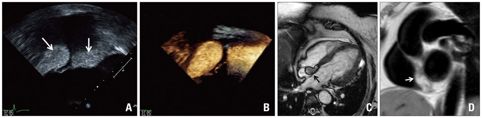
Fig. 1 Two-dimensional (A) and three-dimensional (B) transesophageal echocardiography showed dumbbell-shaped mass with sparing of the fossa ovalis. The mass (arrow) with no contrast enhancement involved on the interatrial septum in cardiac magnetic resonance (C) and showed high signal intensity on T1 weighted image (D).
Reference
-
1. Heyer CM, Kagel T, Lemburg SP, Bauer TT, Nicolas V. Lipomatous hypertrophy of the interatrial septum: a prospective study of incidence, imaging findings, and clinical symptoms. Chest. 2003. 124:2068–2073.2. Shirani J, Roberts WC. Clinical, electrocardiographic and morphologic features of massive fatty deposits ("lipomatous hypertrophy") in the atrial septum. J Am Coll Cardiol. 1993. 22:226–238.3. Silbiger JJ, Bazaz R, Trost B. Lipomatous hypertrophy of the interatrial septum revisited. J Am Soc Echocardiogr. 2010. 23:789–790.
Article
- Full Text Links
-
- Actions
-
Cited
- CITED
-
- Close
- Share
-
- Similar articles
-
- Lipomatous Hypertrophy of Interartiral Septum(LHIS)
- Lipomatous Hypertrophy of the Interatrial Septum: A Case Report
- Embolic Stroke of Undetermined Source Accompanied by Lipomatous Hypertrophy of Interatrial Septum
- A Rare Case of Lipomatous Hypertrophy of the Interventricular Septum
- Lipomatous Hypertrophy of the Interatrial Septum: A 3-Dimensional Transesophageal Echocardiography Appearance
