Endoscopic Resection of Hypopharyngeal Squamous Cell Carcinoma
- Affiliations
-
- 1Gastrointestinal Cancer Center, Soonchunhyang University Hospital, Soonchunhyang University College of Medicine, Seoul, Korea. cjy6695@dreamwiz.com
- KMID: 1805304
- DOI: http://doi.org/10.5946/ce.2013.46.2.189
Abstract
- Hypopharyngeal cancers are often diagnosed at an advanced stage and have a poor prognosis. Even when they are diagnosed at an operable stage, surgery often results in substantial morbidity and decreased patients' quality of life. Although the endoscopic diagnosis of early hypopharyngeal cancer is difficult, recent developments in advanced imaging endoscopy have enabled easier diagnosis of these lesions. Endoscopic resection of early hypopharyngeal cancer is a potential minimally invasive treatment that can preserve the function and quality of life of patients. Reports of this procedure are limited, however. We report a case of hypopharygeal cancer treated with endoscopic resection.
Figure
-
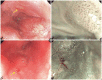
Fig. 1 Endoscopic findings of the hypopharynx. (A) A hyperemic elevated lesion in observed at the right pyriform recess (yellow arrow). (B) Narrow band imaging (NBI) shows a type V irregular intrapapillary capillary loops pattern. (C) A slightly depressed mucosal lesion at the left pyriform recess (yellow arrow) is seen on follow up endoscopy. (D) NBI shows an irregular vascular pattern.
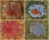
Fig. 2 Gross and pathologic mapping findings of the resected lesions. (A) Specimen from right pyriform recess. A hyperemic elevated lesion measuring approximately 7×3 mm in observed in the center of the specimen. (B) Pathologic mapping shows the lesion in orange with free lateral margins. (C) Specimen from left pyriform recess. A minute mucosal lesion is observed at the upper left hand corner of the specimen. (D) Pathologic mapping show the lesion in orange with free lateral margins.
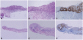
Fig. 3 Microscopic findings of the resected lesion. Top; right pyriform recess. (A, B) The entire epithelial layer is replaced by atypical squamous epithelial cells, (C) accompanied by diffuse strong p53 protein overexpression in their nuclei. Bottom; left pyriform recess. (D, E) The squamous cells in the lower one-third of the epithelial layer is atypical, (F) where p53 protein overexpression can be identified (A, D, H&E stain, ×40; B, E, H&E stain, ×100; C, F, p53, ×100).
Reference
-
1. Shimizu Y, Yamamoto J, Kato M, et al. Endoscopic submucosal dissection for treatment of early stage hypopharyngeal carcinoma. Gastrointest Endosc. 2006; 64:255–259. PMID: 16860078.
Article2. Suzuki H, Saito Y, Oda I, Nonaka S, Nakanishi Y. Feasibility of endoscopic mucosal resection for superficial pharyngeal cancer: a minimally invasive treatment. Endoscopy. 2010; 42:1–7. PMID: 20066588.
Article3. Iizuka T, Kikuchi D, Hoteya S, Yahagi N, Takeda H. Endoscopic submucosal dissection for treatment of mesopharyngeal and hypopharyngeal carcinomas. Endoscopy. 2009; 41:113–117. PMID: 19214888.
Article4. Muto M, Nakane M, Katada C, et al. Squamous cell carcinoma in situ at oropharyngeal and hypopharyngeal mucosal sites. Cancer. 2004; 101:1375–1381. PMID: 15368325.
Article5. Iizuka T, Kikuchi D, Hoteya S, et al. Clinical advantage of endoscopic submucosal dissection over endoscopic mucosal resection for early mesopharyngeal and hypopharyngeal cancers. Endoscopy. 2011; 43:839–843. PMID: 21833903.
Article6. Wang WL, Lee CT, Lee YC, et al. Risk factors for developing synchronous esophageal neoplasia in patients with head and neck cancer. Head Neck. 2011; 33:77–81. PMID: 20848418.
Article7. Inoue H, Honda T, Nagai K, et al. Ultra-high magnification endoscopic observation of carcinoma in situ of the esophagus. Dig Endosc. 1997; 9:16–18.
Article
- Full Text Links
-
- Actions
-
Cited
- CITED
-
- Close
- Share
-
- Similar articles
-
- Diagnosis and Clinical Management of Esophageal Squamous Dysplasia
- Endoscopic Treatment for Esophageal Cancer
- Is it Necessary to Dissect Level I in Laryngeal and Hypopharyngeal Squamous Cell Carcinoma?
- A Case of Early-stage Squamous Cell Carcinoma of the Anal Canal Diagnosed by Endoscopic Mucosal Resection
- Antitumor effects of valdecoxib on hypopharyngeal squamous carcinoma cells
