Imaging Sci Dent.
2014 Jun;44(2):161-164. 10.5624/isd.2014.44.2.161.
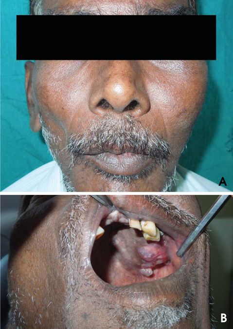
Central mucoepidermoid carcinoma of the maxilla with unusual ground glass appearance and calcifications: A case report
- Affiliations
-
- 1Department of Oral Medicine and Radiology, C.K.S. Teja Dental College, Tirupati, India. sureshomr@gmail.com
- 2Department of Oral Medicine and Radiology, Narayana Dental College and Hospital, Nellore, India.
- KMID: 1799599
- DOI: http://doi.org/10.5624/isd.2014.44.2.161
Abstract
- Mucoepidermoid carcinomas (MECs) arising within the jaws as primary central bony lesions are termed central MECs. Central MECs are extremely rare, comprising 2-3% of all mucoepidermoid carcinomas. We herein report a rare case of central MEC of the maxilla in a 52-year-old male whose plain radiographs showed a "ground glass" pattern and computed tomographic images, a hypodense mass with numerous calcifications. To the best of our knowledge, this is the first report of central MEC showing a "ground glass" appearance.
MeSH Terms
Figure
Reference
-
1. Varma S, Shameena P, Sudha S, Nair RG, Varghese IV. Clear cell variant of intraosseous mucoepidermoid carcinoma: report of a rare entity. J Oral Maxillofac Pathol. 2012; 16:141–144.
Article2. Simon D, Somanathan T, Ramdas K, Pandey M. Central mucoepidermoid carcinoma of mandible - A case report and review of the literature. World J Surg Oncol. 2003; 1:1.3. Tucci R, Matizonkas-Antonio LF, de Carvalhosa AA, Castro PH, Nunes FD, Pinto DD Jr. Central mucoepidermoid carcinoma: report of a case with 11 years' evolution and peculiar macroscopical and clinical characteristics. Med Oral Patol Oral Cir Bucal. 2009; 14:E283–E286.4. Rabinov JD. Imaging of salivary gland pathology. Radiol Clin North Am. 2000; 38:1047–1057.
Article5. Browand BC, Waldron CA. Central mucoepidermoid tumors of the jaws. Report of nine cases and review of the literature. Oral Surg Oral Med Oral Pathol. 1975; 40:631–643.6. Brookstone MS, Huvos AG. Central salivary gland tumors of the maxilla and mandible: a clinicopathologic study of 11 cases with an analysis of the literature. J Oral Maxillofac Surg. 1992; 50:229–236.
Article7. do Prado RF, Lima CF, Pontes HA, Almeida JD, Cabral LA, Carvalho YR, et al. Calcifications in a clear cell mucoepidermoid carcinoma: a case report with histological and immunohistochemical findings. Oral Surg Oral Med Oral Pathol Oral Radiol Endod. 2007; 104:e40–e44.
Article8. Sherin S, Sherin N, Thomas V, Kumar N, Sharafuddeen KP. Central mucoepidermoid carcinoma of maxilla with radiographic appearance of mixed radiopaque-radiolucent lesion: a case report. Dentomaxillofac Radiol. 2011; 40:463–465.
Article9. Kurabayashi T, Ida M, Yoshino N, Sasaki T, Ishii J, Ueda M. Differential diagnosis of tumours of the minor salivary glands of the palate by computed tomography. Dentomaxillofac Radiol. 1997; 26:16–21.
Article10. Yoon JH, Ahn SG, Kim SG, Kim J. Calcifications in a clear cell mucoepidermoid carcinoma of the hard palate. Int J Oral Maxillofac Surg. 2005; 34:927–929.
Article
- Full Text Links
- Actions
-
Cited
- CITED
-
- Close
- Share
- Similar articles
-
- Mucoepidermoid carcinoma in the mandible : review of a case
- Central Mucoepidermoid Carcinoma of the Mandible: Case Report
- A Case of Conjunctival Mucoepidermoid Carcinoma with invioving Cornea
- A Case of Solitary Cutaneous reticulohistiocytoma
- Cytopathology of Metastatic Mucoepidermoid Carcioma of the Lung


