Outcomes of open versus closed treatment in the management of mandibular subcondylar fractures
- Affiliations
-
- 1Department of Oral and Maxillofacial Surgery, Gachon University Gil Medical Center, Incheon, Korea. face@gilhospital.com
- KMID: 1799564
- DOI: http://doi.org/10.5125/jkaoms.2014.40.6.297
Abstract
OBJECTIVES
To compare the clinical and radiological outcomes after closed reduction (CR) and open reduction and internal fixation (ORIF) in the management of subcondylar fractures.
MATERIALS AND METHODS
Forty-eight patients presenting with subcondylar fracture between January 2010 and March 2013 were evaluated retrospectively. Fifteen patients were treated with CR and 33 patients with ORIF. The clinical and radiologic parameters were evaluated during follow-up (mean, 7.06 months; range, 3 to 36 months).
RESULTS
In the CR group, no patients had any problems with regard to the clinical parameters. The average period of maxillomandibular fixation (MMF) was 5.47 days. The preoperative average tangential angulation of the fractured fragment was 3.67degrees, and loss of ramus height was 2.44 mm. In the ORIF group, no clinical problems were observed, and the average period of MMF was 6.33 days. The preoperative average tangential angulation of the subcondylar fragment was 8.66degrees, and loss of ramus height was 3.61 mm.
CONCLUSION
CR provided satisfactory clinical results, though ORIF provided more accurate reduction of the fractured fragment. So there is no distinct displacement of fractured fragment, CR should be selected than ORIF because of no need for surgery.
Figure
-
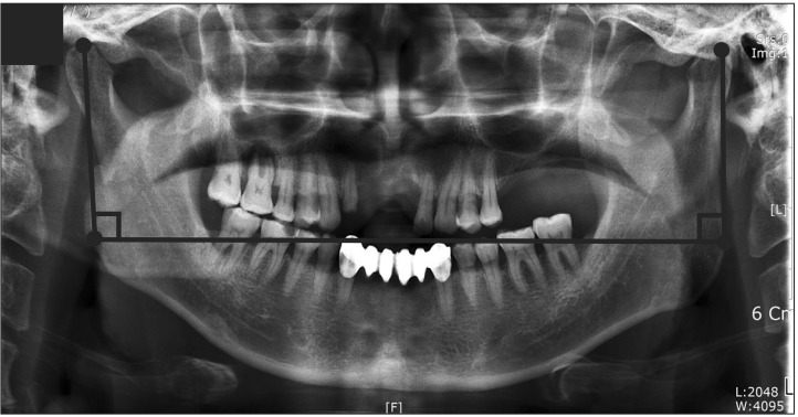
Fig. 1 Illustration showing the method by which loss of ramus height was measured on the panoramic view. A reference line was drawn through both gonial angles. A perpendicular line between the most superior point of the condyle and the reference line was drawn on the panoramic radiograph. The difference between the non-fractured and fractured side was used as a measure of difference in ramus length.
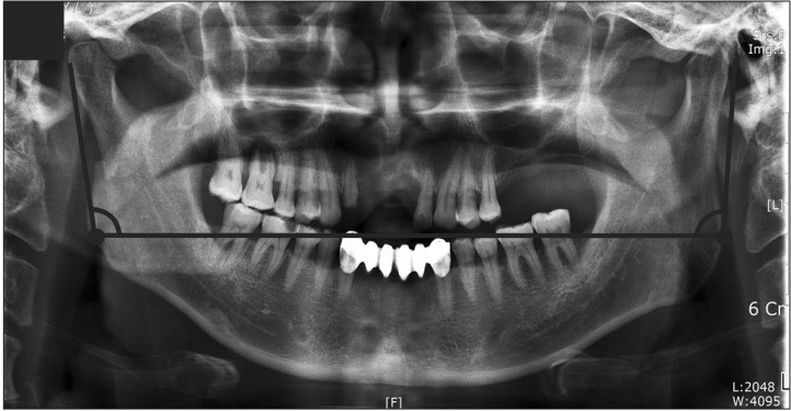
Fig. 2 Illustration showing the method by which tangential displacement was quantified on the panoramic view. A reference line was drawn through both gonial angles, and another line was drawn tangential to the posterior border of the condylar process on each side. The angle between the intersection of the tangent and the condylar process was calculated. The difference in this angle between the non-fractured and fractured sides was used as a measurement of tangential angulation.
Cited by 1 articles
-
New protocol for simplified reduction and fixation of subcondylar fractures of the mandible: a technical note
Saurabh Mohandas Kamat, Vikas Dhupar, Francis Akkara
J Korean Assoc Oral Maxillofac Surg. 2021;47(5):403-406. doi: 10.5125/jkaoms.2021.47.5.403.
Reference
-
1. Lee SC, Kim YG, Ryu DM, Lee BS, Yoon OB, Jin TH. A clinical and statistical study of condylar fracture of mandible. J Korean Assoc Oral Maxillofac Surg. 1998; 24:326–329.2. Lindahl L. Condylar fractures of the mandible. I. Classification and relation to age, occlusion, and concomitant injuries of teeth and teeth-supporting structures, and fractures of the mandibular body. Int J Oral Surg. 1977; 6:12–21. PMID: 402318.3. Palmieri C, Ellis E 3rd, Throckmorton G. Mandibular motion after closed and open treatment of unilateral mandibular condylar process fractures. J Oral Maxillofac Surg. 1999; 57:764–775. PMID: 10416622.
Article4. Brandt MT, Haug RH. Open versus closed reduction of adult mandibular condyle fractures: a review of the literature regarding the evolution of current thoughts on management. J Oral Maxillofac Surg. 2003; 61:1324–1332. PMID: 14613090.
Article5. Suzuki T, Kawamura H, Kasahara T, Nagasaka H. Resorbable poly-L-lactide plates and screws for the treatment of mandibular condylar process fractures: a clinical and radiologic follow-up study. J Oral Maxillofac Surg. 2004; 62:919–924. PMID: 15278854.
Article6. Iizuka T, Lädrach K, Geering AH, Raveh J. Open reduction without fixation of dislocated condylar process fractures: long-term clinical and radiologic analysis. J Oral Maxillofac Surg. 1998; 56:553–561. PMID: 9590337.
Article7. Undt G, Kermer C, Rasse M, Sinko K, Ewers R. Transoral miniplate osteosynthesis of condylar neck fractures. Oral Surg Oral Med Oral Pathol Oral Radiol Endod. 1999; 88:534–543. PMID: 10556746.
Article8. Baker AW, McMahon J, Moos KF. Current consensus on the management of fractures of the mandibular condyle. A method by questionnaire. Int J Oral Maxillofac Surg. 1998; 27:258–266. PMID: 9698171.
- Full Text Links
-
- Actions
-
Cited
- CITED
-
- Close
- Share
-
- Similar articles
-
- Comparison Study of Open Reduction and Closed Reduction in Treatment of Mandibular Subcondylar Fractures
- Clinical Applications of Endoscopic-Assisted Open Reduction and Internal Fixation of Subcondylar Fractures
- Intraoral Open Reduction of Mandibular Subcondylar Fractures using Kirschner Wire
- Surgical Management of a Mandible Subcondylar Fracture
- Current Concepts in the Mandibular Condyle Fracture Management Part II: Open Reduction Versus Closed Reduction
