Yonsei Med J.
2013 Nov;54(6):1484-1490. 10.3349/ymj.2013.54.6.1484.
The Relation between Inferior Mesenteric Vein Ligation and Collateral Vessels to Splenic Flexure: Anatomical Landmarks, Technical Precautions and Clinical Significance
- Affiliations
-
- 1Section of Colon and Rectal Surgery, Department of Surgery, Yonsei University College of Medicine, Seoul, Korea. namkyuk@yuhs.ac, sss_Allah@hotmail.com
- KMID: 1798147
- DOI: http://doi.org/10.3349/ymj.2013.54.6.1484
Abstract
- PURPOSE
Our aim to assess clinical significance of the relation between inferior mesenteric vein ligation and collateral blood supply (meandering mesenteric artery) to the splenic flexure with elaboration more in anatomical landmarks and technical tips.
MATERIALS AND METHODS
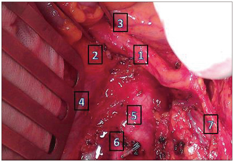
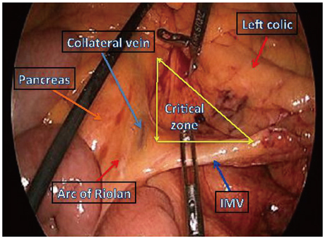
We review the literature regarding the significance of the collateral vessels around inferior mesenteric vein (IMV) root and provide our prospective operative findings, anatomical landmarks and technical tips. We analyzed the incidence and pattern of anatomic variation of collateral vessels around the IMV.
RESULTS
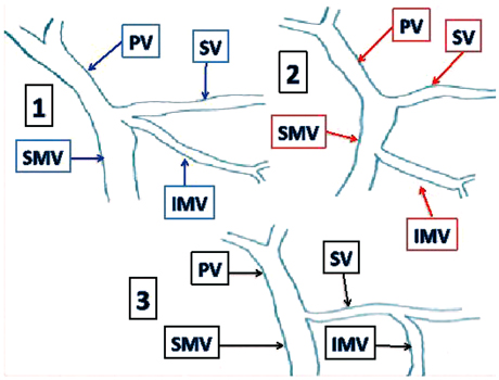
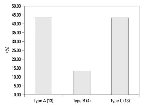
A total of 30 consecutive patients have been prospectively observed in a period between June 25-2012 and September 7-2012. Nineteen males and eleven females with mean age of 63 years. Major colorectal procedures were included. There were three anatomical types proposed, based on the relation between IMV and the collateral vessel. Type A and B in which either the collateral vessel crosses or runs close to the IMV with incidence of 43.3% and 13.3%, respectively, whereas type C is present in 43.3%. There was no definitive relation between the artery and vein. No intra or postoperative ischemic events were reported.
CONCLUSION
During IMV ligation, inadvertent ligation of Arc of Riolan or meandering mesenteric artery around the IMV root "in type A&B" might result in compromised blood supply to the left colon, congestion, ischemia and different level of colitis or anastomotic dehiscence. Therefore, careful dissection and skeletonization at the IMV root "before ligation if necessary" is mandatory to preserve the collateral vessel for the watershed area and to avoid further injury.
MeSH Terms
Figure
Reference
-
1. Park MG, Hur H, Min BS, Lee KY, Kim NK. Colonic ischemia following surgery for sigmoid colon and rectal cancer: a study of 10 cases and a review of the literature. Int J Colorectal Dis. 2012; 27:671–675.
Article2. Netter Frank H.. Essentials Atlas of Human Anatomy. ISBN 0-914168-18-5 Copyright 1987.3. Walker TG. Mesenteric vasculature and collateral pathways. Semin Intervent Radiol. 2009; 26:167–174.
Article4. Sakorafas GH, Zouros E, Peros G. Applied vascular anatomy of the colon and rectum: clinical implications for the surgical oncologist. Surg Oncol. 2006; 15:243–255.
Article5. Michels NA, Siddharth P, Kornblith PL, Parke WW. The variant blood supply to the descending colon, rectosigmoid and rectum based on 400 dissections. Its importance in regional resections: a review of medical literature. Dis Colon Rectum. 1965; 8:251–278.
Article6. Kachlik D, Baca V. Macroscopic and microscopic intermesenteric communications. Biomed Pap Med Fac Univ Palacky Olomouc Czech Repub. 2006; 150:121–124.
Article7. Gourley EJ, Gering SA. The meandering mesenteric artery: a historic review and surgical implications. Dis Colon Rectum. 2005; 48:996–1000.
Article8. Rosenblum JD, Boyle CM, Schwartz LB. The mesenteric circulation. Anatomy and physiology. Surg Clin North Am. 1997; 77:289–306.9. Horton KM, Fishman EK. Volume-rendered 3D CT of the mesenteric vasculature: normal anatomy, anatomic variants, and pathologic conditions. Radiographics. 2002; 22:161–172.
Article10. Bernstein WC, Bernstein EF. Ischemic ulcerative colitis following inferior mesenteric arterial ligation. Dis Colon Rectum. 1963; 6:54–61.
Article11. Moskowitz M, Zimmerman H, Felson B. The meandering mesenteric artery of the colon. Am J Roentgenol Radium Ther Nucl Med. 1964; 92:1088–1099.12. Douard R, Chevallier JM, Delmas V, Cugnenc PH. Clinical interest of digestive arterial trunk anastomoses. Surg Radiol Anat. 2006; 28:219–227.
Article13. Pikkieff H. Über die Blutversorgung des Dickendarms. Zschr Anat Entw. 1931; 96:658–679.14. Lin PH, Chaikof EL. Embryology, anatomy, and surgical exposure of the great abdominal vessels. Surg Clin North Am. 2000; 80:417–433.
Article15. Baden JG, Racy DJ, Grist TM. Contrast-enhanced three-dimensional magnetic resonance angiography of the mesenteric vasculature. J Magn Reson Imaging. 1999; 10:369–375.
Article16. Meaney JF. Non-invasive evaluation of the visceral arteries with magnetic resonance angiography. Eur Radiol. 1999; 9:1267–1276.
Article17. Shirkhoda A, Konez O, Shetty AN, Bis KG, Ellwood RA, Kirsch MJ. Mesenteric circulation: three-dimensional MR angiography with a gadolinium-enhanced multiecho gradient-echo technique. Radiology. 1997; 202:257–261.
Article18. Therasse E, Soulez G, Roy P, Gauvin A, Oliva VL, Carrier R, et al. Lower extremity: nonstepping digital angiography with photostimulable imaging plates versus conventional angiography. Radiology. 1998; 207:695–703.
Article19. Grinnell RS, Hiatt RB. Ligation of the interior mesenteric artery at the aorta in resections for carcinoma of the sigmoid and rectum. Surg Gynecol Obstet. 1952; 94:526–534.20. Goligher JC. The adequacy of the marginal blood-supply to the left colon after high ligation of the inferior mesenteric artery during excision of the rectum. Br J Surg. 1954; 41:351–353.
Article21. Morgan CN, Griffiths JD. High ligation of the inferior mesenteric artery during operations for carcinoma of the distal colon and rectum. Surg Gynecol Obstet. 1959; 108:641–650.22. Feldman M, Scharschmidt BF, Sleisenger MH. Sleisenger and Fordtran's: Gastrointestinal and liver Disease: Pathophysiology/Diagnosis/Management. 9th ed. Philadelphia: Saunders/Elsevier;2010.23. Sakanoue Y, Kusunoki M, Shoji Y, Kusuhara K, Yamamura T, Utsunomiya J. Passage of a colon 'cast' after anoabdominal rectal resection. Report of a case. Dis Colon Rectum. 1990; 33:1044–1046.24. Erguney S, Yavuz N, Ersoy YE, Teksoz S, Selcuk D, Ogut G, et al. Passage of "colonic cast" after colorectal surgery: report of four cases and review of the literature. J Gastrointest Surg. 2007; 11:1045–1051.
Article25. Parc R, Cugnenc PH, Levy E, Huguet C, Loygue J. [Early post-operative complications in intestinal resections followed with colo-colitic or recto-colitic anastomoses. Clinical and biological manifestations of anastomotic complications. Therapeutic results about 523 cases (author's transl)]. Ann Chir. 1981; 35:69–82.26. Einstein AJ, McLaughlin MA, Lipman HI, Sanz J, Rajagopalan S. Images in vascular medicine the Arc of Riolan: diagnosis by magnetic resonance angiography. Vasc Med. 2005; 10:239.27. Cima RR, Bilings B. Strategies to avoid 3 common problems in colorectal surgery. Contemp Surg. 2008; 64:120–125.28. Seike K, Koda K, Saito N, Oda K, Kosugi C, Shimizu K, et al. Laser Doppler assessment of the influence of division at the root of the inferior mesenteric artery on anastomotic blood flow in rectosigmoid cancer surgery. Int J Colorectal Dis. 2007; 22:689–697.
Article
- Full Text Links
- Actions
-
Cited
- CITED
-
- Close
- Share
- Similar articles
-
- Laparoscopic Splenic Flexure Mobilization Using a Medial Approach
- Splenoportography in portal hypertension
- An analysis of splenoportographic findings in portal hypertension
- Blood flow volume difference (P-SS) between the portal vein and thesum of splenic vein and superior mesenteric vein in portal hypertension
- A Case of the Inferior Mesenteric Artery Arising from the Superior Mesenteric Artery in a Korean Woman


