Diagnostic Imaging Utilization in Cases of Acute Appendicitis: Multi-Center Experience
- Affiliations
-
- 1Department of Radiology, Seoul National University Bundang Hospital, Seongnam, Korea. pjihoon79@gmail.com
- KMID: 1794612
- DOI: http://doi.org/10.3346/jkms.2014.29.9.1308
Abstract
- The purpose of this cross-sectional study was to measure imaging utilization rates and the negative appendectomy rate (NAR) in metropolitan Seoul, Korea. The study included 2321 adolescents and adults (> or =15 yr; median [interquartile range] age, 37 [27-50] yr; 46.7% female) undergoing appendectomy in 2011 at eight tertiary and three secondary hospitals. Imaging utilization rate was 99.7% (95% confidence interval, 99.4%-99.9%). CT and ultrasonography utilization rates as an initial imaging modality were 93.1% (92.0%-94.1%), and 6.5% (5.6%-7.6%), respectively. The NAR in patients undergoing CT only, complementary ultrasonography following CT, ultrasonography only, and complementary CT following ultrasonography were 3.3% (2.6%-4.1%), 27% (14%-44%), 9% (4%-16%), and 8% (2%-20%), respectively. The use of ultrasonography instead of CT as the initial imaging modality was significantly associated with higher NAR (adjusted odds ratio [AOR], 2.28 [1.22-4.27]; risk difference, 4.4 [0-8.8] percentage points), however, the population attributable risk was 0.3 [0-0.6] percentage points. We observed a very high CT utilization rate and a low NAR in metropolitan Seoul. Although the use of CT was significantly associated with the lower NAR, CT utilization rate already has reached the level that increase in CT utilization from the status quo would hardly decrease the NAR further.
MeSH Terms
Figure
-
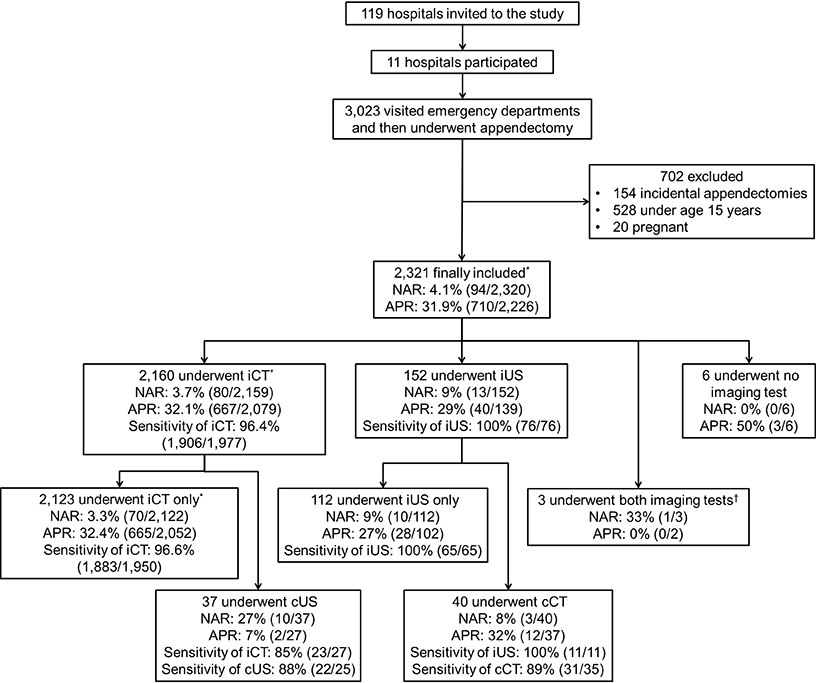
Fig. 1 Study flow diagram. APR indicates appendiceal perforation rate; cCT, complementary computed tomography; cUS, complementary ultrasonography; iCT, initial computed tomography; iUS, initial ultrasonography; NAR, negative appendectomy rate. Cases with unavailable computed tomography (n = 106) or ultrasonography (n = 67) reports were not included when calculating the sensitivities. When calculating the sensitivities, indeterminate results were counted as a positive diagnosis. *Includes one patient with a missing pathology report; †The order of the two imaging tests was unclear.
Cited by 4 articles
-
Structured Reporting versus Free-Text Reporting for Appendiceal Computed Tomography in Adolescents and Young Adults: Preference Survey of 594 Referring Physicians, Surgeons, and Radiologists from 20 Hospitals
Sung Bin Park, Min-Jeong Kim, Yousun Ko, Ji Ye Sim, Hyuk Jung Kim, Kyoung Ho Lee,
Korean J Radiol. 2019;20(2):246-255. doi: 10.3348/kjr.2018.0109.Using 2-mSv Appendiceal CT in Usual Practice for Adolescents and Young Adults: Willingness Survey of 579 Radiologists, Emergency Physicians, and Surgeons from 20 Hospitals
Hyuk Jung Kim, Kyoung Ho Lee, Min-Jeong Kim, Sung Bin Park, Yousun Ko,
Korean J Radiol. 2020;21(1):68-76. doi: 10.3348/kjr.2019.0010.Meaningful standard of reference for appendiceal perforation: pathology, surgery, or both?
Hyuk Jung Kim, Mi Sung Kim, Ji Hoon Park, Soyeon Ahn, Yousun Ko, Soon-Young Song, Ji Young Woo, Kyoung Ho Lee,
Ann Surg Treat Res. 2017;93(2):88-97. doi: 10.4174/astr.2017.93.2.88.Central Image Archiving and Management System for Multicenter Clinical Studies: Lessons from Low-dose CT for Appendicitis Trial
Yousun Ko, Jin Woo Choi, Dong Hyun Kim, Kyong Joon Lee, Sang Soo Shin, Ji Young Woo, Seong Whi Cho, Bong Soo Kim, Kyoung Ho Lee
J Korean Soc Radiol. 2017;76(3):165-172. doi: 10.3348/jksr.2017.76.3.165.
Reference
-
1. Coursey CA, Nelson RC, Patel MB, Cochran C, Dodd LG, Delong DM, Beam CA, Vaslef S. Making the diagnosis of acute appendicitis: do more preoperative CT scans mean fewer negative appendectomies? a 10-year study. Radiology. 2010; 254:460–468.2. Cuschieri J, Florence M, Flum DR, Jurkovich GJ, Lin P, Steele SR, Symons RG, Thirlby R. Negative appendectomy and imaging accuracy in the Washington State Surgical Care and Outcomes Assessment Program. Ann Surg. 2008; 248:557–563.3. Drake FT, Florence MG, Johnson MG, Jurkovich GJ, Kwon S, Schmidt Z, Thirlby RC, Flum DR. SCOAP Collaborative. Progress in the diagnosis of appendicitis: a report from Washington State's Surgical Care and Outcomes Assessment Program. Ann Surg. 2012; 256:586–594.4. Raja AS, Wright C, Sodickson AD, Zane RD, Schiff GD, Hanson R, Baeyens PF, Khorasani R. Negative appendectomy rate in the era of CT: an 18-year perspective. Radiology. 2010; 256:460–465.5. Raman SS, Osuagwu FC, Kadell B, Cryer H, Sayre J, Lu DS. Effect of CT on false positive diagnosis of appendicitis and perforation. N Engl J Med. 2008; 358:972–973.6. Rao PM, Rhea JT, Rattner DW, Venus LG, Novelline RA. Introduction of appendiceal CT: impact on negative appendectomy and appendiceal perforation rates. Ann Surg. 1999; 229:344–349.7. Rhea JT, Halpern EF, Ptak T, Lawrason JN, Sacknoff R, Novelline RA. The status of appendiceal CT in an urban medical center 5 years after its introduction: experience with 753 patients. AJR Am J Roentgenol. 2005; 184:1802–1808.8. Sugihara K, Muto T, Morioka Y, Asano A, Yamamoto T. Diverticular disease of the colon in Japan: a review of 615 cases. Dis Colon Rectum. 1984; 27:531–537.9. Kim K, Kim YH, Kim SY, Kim S, Lee YJ, Kim KP, Lee HS, Ahn S, Kim T, Hwang SS, et al. Low-dose abdominal CT for evaluating suspected appendicitis. N Engl J Med. 2012; 366:1596–1605.10. Pooler BD, Lawrence EM, Pickhardt PJ. Alternative diagnoses to suspected appendicitis at CT. Radiology. 2012; 265:733–742.11. Horton MD, Counter SF, Florence MG, Hart MJ. A prospective trial of computed tomography and ultrasonography for diagnosing appendicitis in the atypical patient. Am J Surg. 2000; 179:379–381.12. Pickuth D, Heywang-Köbrunner SH, Spielmann RP. Suspected acute appendicitis: is ultrasonography or computed tomography the preferred imaging technique? Eur J Surg. 2000; 166:315–319.13. Balthazar EJ, Birnbaum BA, Yee J, Megibow AJ, Roshkow J, Gray C. Acute appendicitis: CT and US correlation in 100 patients. Radiology. 1994; 190:31–35.14. Van Randen A, Bipat S, Zwinderman AH, Ubbink DT, Stoker J, Boermeester MA. Acute appendicitis: meta-analysis of diagnostic performance of CT and graded compression US related to prevalence of disease. Radiology. 2008; 249:97–106.15. Terasawa T, Blackmore CC, Bent S, Kohlwes RJ. Systematic review: computed tomography and ultrasonography to detect acute appendicitis in adults and adolescents. Ann Intern Med. 2004; 141:537–546.16. Lee KH, Kim YH, Hahn S, Lee KW, Lee HJ, Kim TJ, Kang SB, Shin JH, Park BJ. Added value of coronal reformations for duty radiologists and for referring physicians or surgeons in the CT diagnosis of acute appendicitis. Korean J Radiol. 2006; 7:87–96.17. Ahn S. LOCAT group. LOCAT (low-dose computed tomography for appendicitis trial) comparing clinical outcomes following low- vs standard-dose computed tomography as the first-line imaging test in adolescents and young adults with suspected acute appendicitis: study protocol for a randomized controlled trial. Trials. 2014; 15:28.18. Health Insurance Review & Assessment Service of Korea. Hospital survey. accessed on 21 May 2013. Available at http://www.hira.or.kr/main.do.19. National Health Insurance Service. 2011 statistics on major surgeries in Korea. accessed on 20 May 2013. Available at http://www.bokjiro.go.kr/data/statusView.do?board_sid=297&data_sid=5999111.20. Saito JM, Yan Y, Evashwick TW, Warner BW, Tarr PI. Use and accuracy of diagnostic imaging by hospital type in pediatric appendicitis. Pediatrics. 2013; 131:e37–e44.21. Rosai J. Appendix. In : Rosai J, editor. Rosai and Ackerman's surgical pathology. 9th ed. Edinburgh: Mosby;2004. p. 758–759.22. Fenoglio-Preiser CM, Noffsinger AE, Stemmermann GN, Lantz PE, Isaacson PG. Nonneoplastic diseases of the appendix. In : Fenoglio-Preiser CM, Noffsinger AE, Stemmermann GN, Lantz PE, Isaacson PG, editors. Gastrointestinal pathology: an atlas and text. 3rd ed. Philadelphia: Lippincott Williams & Wilkins;2008. p. 504–505.23. Lee KH, Lee HS, Park SH, Bajpai V, Choi YS, Kang SB, Kim KJ, Kim YH. Appendiceal diverticulitis: diagnosis and differentiation from usual acute appendicitis using computed tomography. J Comput Assist Tomogr. 2007; 31:763–769.24. Phillips BJ, Perry CW. Appendiceal diverticulitis. Mayo Clin Proc. 1999; 74:890–892.25. Flum DR, Morris A, Koepsell T, Dellinger EP. Has misdiagnosis of appendicitis decreased over time? a population-based analysis. JAMA. 2001; 286:1748–1753.26. Sim JY, Kim HJ, Yeon JW, Suh BS, Kim KH, Ha YR, Paik SY. Added value of ultrasound re-evaluation for patients with equivocal CT findings of acute appendicitis: a preliminary study. Eur Radiol. 2013; 23:1882–1890.27. Greenland S. Applications of stratified analysis methods. In : Rothman KJ, Greenland S, Lash TL, editors. Modern epidemiology. 3rd ed. Philadelphia: Lippincott Williams & Wilkins;2008. p. 283–302.28. Wikipedia. Population density. accessed on 20 January 2014. Available at http://en.wikipedia.org/wiki/Population_density.29. Paulson EK, Kalady MF, Pappas TN. Clinical practice. Suspected appendicitis. N Engl J Med. 2003; 348:236–242.30. Birnbaum BA, Wilson SR. Appendicitis at the millennium. Radiology. 2000; 215:337–348.31. National Council on Radiation Protection and Measurements. Ionizing radiation exposure of the population of the United States: NCRP Report No. 160. Bethesda: National Council on Radiation Protection and Measurements;2009.32. Brix G, Nagel HD, Stamm G, Veit R, Lechel U, Griebel J, Galanski M. Radiation exposure in multi-slice versus single-slice spiral CT: results of a nationwide survey. Eur Radiol. 2003; 13:1979–1991.33. Hart D, Wall BF, Hiller MC, Shrimpton PC. Frequency and collective dose for medical and dental X-ray examinations in the UK, 2008. Chilton: Health Protection Agency;2010.34. Smith-Bindman R, Lipson J, Marcus R, Kim KP, Mahesh M, Gould R, Berrington de González A, Miglioretti DL. Radiation dose associated with common computed tomography examinations and the associated lifetime attributable risk of cancer. Arch Intern Med. 2009; 169:2078–2086.35. Preston DL, Ron E, Tokuoka S, Funamoto S, Nishi N, Soda M, Mabuchi K, Kodama K. Solid cancer incidence in atomic bomb survivors: 1958-1998. Radiat Res. 2007; 168:1–64.36. Brenner DJ, Hall EJ. Computed tomography: an increasing source of radiation exposure. N Engl J Med. 2007; 357:2277–2284.37. National Academy of Sciences. Health risks from exposure to low levels of ionizing radiation: biological effects of ionizing radiation VII phase 2. Washington, D.C.: National Research Council of the National Academies;2005.38. Owings MF, Kozak LJ. Ambulatory and inpatient procedures in the United States, 1996. Vital Health Stat 13. 1998; (139):1–119.39. Pickhardt PJ, Lawrence EM, Pooler BD, Bruce RJ. Diagnostic performance of multidetector computed tomography for suspected acute appendicitis. Ann Intern Med. 2011; 154:789–796.40. Guite KM, Hinshaw JL, Ranallo FN, Lindstrom MJ, Lee FT Jr. Ionizing radiation in abdominal CT: unindicated multiphase scans are an important source of medically unnecessary exposure. J Am Coll Radiol. 2011; 8:756–761.
- Full Text Links
-
- Actions
-
Cited
- CITED
-
- Close
- Share
-
- Similar articles
-
- Appendicitis – Is a Clinical Diagnosis Enough?
- Radiologic diagnosis of acute appendicitis
- Three Cases of Appendicitis Diagnosed by Colonoscopy in Patients with Atypical Presentations
- Multi-Detector CT Findings of Typical and Atypical Appendicitis: A Pictorial Essay
- Magnetic resonance imaging as the first diagnostic imaging modality for pregnant women with suspected acute appendicitis
