J Korean Med Sci.
2009 Feb;24(1):126-131. 10.3346/jkms.2009.24.1.126.
Congenital Middle Ear Cholesteatoma in Children; Retrospective Review of 35 Cases
- Affiliations
-
- 1Department of Otolaryngology Head & Neck Surgery, College of Medicine, the Catholic University of Korea, Seoul, Korea. swyeo@catholic.ac.kr
- KMID: 1794418
- DOI: http://doi.org/10.3346/jkms.2009.24.1.126
Abstract
- Congenital middle ear cholesteatoma (CMEC) is a rare disease entity in otolaryngology. However, we try to assess the characteristic features and recurrences of CMEC in pediatric patients according to stages, and to determine the value of preoperative computed tomography (CT) scan. Retrospective review of 35 cases of CMEC under the age of 15 yr that had been treated at the tertiary referral center from 1995 through 2006. The main outcome measures were CT findings, surgical findings, recurrence rate and hearing assessment. Preoperative CT scan accurately predicted the extent of the cholesteatoma seen during surgery in 30/35 (85.7%). The recurrence rate of CMEC was 5.7% (2/35) and all of recurred cases were stage IV. In recurred cases, cholesteatomas were extended to sinus tympani and facial recess at revisional operation as well as initial operation. So we concluded that preoperative CT scan is essential in defining the extent of existing pathology. The intraoperative CMEC extension and location influence the outcome of surgery. In the higher stages, careful eradication of disease, particularly in the region of sinus tympani and facial recess is recommended.
MeSH Terms
Figure
-
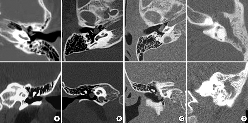
Fig. 1 Stage according to CT findings. (A) Stage I. Cholesteatoma occupies a single quadrant without ossicular extension. (B) Stage II. Cholesteatoma occupies at least two quadrants without mastoid extension. (C) Stage III. Cholesteatoma involve the ossicle. (D) Stage IV. Cholesteatoma extends into the mastoid.
Reference
-
1. McDonald TJ, Cody DT, Ryan RE Jr. Congenital cholesteatoma of the ear. Ann Otol Rhinol Laryngol. 1984. 93:637–640.
Article2. Friedberg J. Congenital cholesteatoma. Laryngoscope. 1994. 104:1–24.
Article3. Aimi K. Role of the tympanic ring in the pathogenesis of congenital cholesteatoma. Laryngoscope. 1983. 93:1140–1146.
Article4. Michaels L. An epidermoid formation in the developing middle ear: possible source of cholesteatoma. J Otolaryngol. 1986. 15:169–174.5. Koltai PJ, Nelson M, Castellon RJ, Garabedian EN, Triglia JM, Roman S, Roger G. The natural history of congenital cholesteatoma. Arch Otolaryngol Head Neck Surg. 2002. 128:804–809.
Article6. Nelson M, Roger G, Koltai PJ, Garabedian EN, Triglia JM, Roman S, Castellon RJ, Hammel JP. Congenital cholesteatoma: classification, management, outcome. Arch Otolaryngol Head Neck Surg. 2002. 128:810–814.7. Potsic WP, Samadi DS, Marsh RR, Wetmore RF. A staging system for congenital cholesteatoma. Arch Otolaryngol Head Neck Surg. 2002. 128:1009–1012.
Article8. Levenson MJ, Michaels L, Parisier SC. Congenital cholesteatomas of the middle ear in children: origin and management. Otolaryngol Clin north Am. 1989. 22:941–954.
Article9. House HP. An apparent primary cholesteatoma, case report. Laryngoscope. 1953. 63:712–713.10. Derlacki EL, Clemis JD. Congenital cholesteatoma of the middle ear and mastoid. Ann Otol Rhinol Laryngol. 1965. 74:706–727.11. Zappia JJ, Wiet RJ. Congenital cholesteatoma. Arch Otolaryngol Head Neck Surg. 1995. 121:19–22.
Article12. House JW, Sheehy JL. Cholesteatoma with intact tympanic membrane: A report of 41 cases. Laryngoscope. 1980. 90:70–76.
Article13. Karmarkar S, Bhatia S, Khashaba A, Saleh E, Russo A, Sanna M. Congenital cholesteatomas of the middle ear: a different experience. Am J Otol. 1996. 17:288–292.14. Kazahaya K, Potsic WP. Congenital cholesteatoma. Curr Opin Otolaryngol Head Neck Surg. 2004. 12:398–403.
Article15. Rizer FM, Luxford WM. The management of congenital cholesteatoma: surgical results of 42 cases. Laryngoscope. 1988. 98:254–256.16. Ueda H, Nakashima T, Nakata S. Surgical strategy for cholesteatoma in children. Auris Nasus Larynx. 2001. 28:125–129.
Article17. Gocmen H, Kilic R, Ozdek A, Kizilkaya Z, Safak MA, Samim E. Surgical treatment of cholesteatoma in children. Int J Pediatr Otorhinolaryngol. 2003. 67:867–872.18. Darrouzet V, Duclos JY, Portmann D, Bebear JP. Congenital middle ear cholesteatomas in children: our experience in 34 cases. Otolaryngol Head Neck Surg. 2002. 126:34–40.
Article19. El-Bitar MA, Choi SS, Emamian SA, Vezina LG. Congenital middle ear cholesteatoma: need for early recognition-role of computed tomography scan. Int J Pediatr Otorhinolaryngol. 2003. 67:231–235.
Article20. Kim CN, Chung SM, Kim SM, Kim YJ, Park MH, Ju MS. Study of the correlation with the temporal bone CT and operative findings in chronic otitis media with cholesteatoma. Korean J Otolaryngol. 1993. 36:313–320.
- Full Text Links
-
- Actions
-
Cited
- CITED
-
- Close
- Share
-
- Similar articles
-
- A Case of Two Isolated Congenital Cholesteatomas Presented in Middle Ear Cavity
- A Case of Congenital Cholesteatoma : Combined with Ossicular Anomaly
- A Case of Bilateral Congenital Middle Ear Cholesteatoma
- A Case of Simultaneous Presentation of Closed and Open Type of Congenital Cholesteatoma
- A Case of Intratympanic Membrane Congenital Cholesteatoma
