Primary Squamous Cell Carcinoma in the Testis: A Case Report
- Affiliations
-
- 1Department of Pathology, Gachon University of Medicine and Science, Gil Medical Center, Incheon, Korea. syha@gilhospital.com
- 2Department of Urology, Gachon University of Medicine and Science, Gil Medical Center, Incheon, Korea.
- 3Department of Forensic Medicine, National Institute of Scientific Investigation, Seoul, Korea.
- KMID: 1792938
- DOI: http://doi.org/10.3346/jkms.2010.25.4.634
Abstract
- A 51-yr-old man presented with an enlarged right testis for two months. The radically resected testis showed a relatively well-circumscribed ovoid mass, nearly replacing the normal architecture with central cystic changes. Microscopically, the mass was composed of ovoid shaped tumor cells of a moderately differentiated squamous cell carcinoma (SCC). The central portion of the mass was filled with well-formed laminated keratinous materials and the remnant cavity lined by dysplastic squamous epithelium, indicated SCC may be derived from an epidermal cyst. SCC is among the most common types of neoplasm afflicting human beings, but it is rare in the testis. To our knowledge, this is the second report of the testicular squamous cell carcinoma occurring in a patient without other primary tumors, and the firstly reported case in Korea.
Keyword
MeSH Terms
Figure
-

Fig. 1 Abdominal computed tomography shows a well-circumscribed oval shaped mass with heterogeneous internal density (arrow).
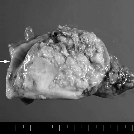
Fig. 2 Photograph of the resected specimen shows that an oval shaped mass nearly replaces the testis. Arrow indicates a hydrocele.

Fig. 3 Histopathological findings of the testis. (A) Nests of squamous cell carcinoma show wall infiltration (H&E stain, ×200). (B) High magnification of neoplastic squamous cells shows orangeophilic cytoplasm and intercellular bridges (H&E stain, ×400). (C) Central cyst, probably epidermal cyst, is filled with laminated linear keratinous materials (×40). Inset indicates foreign body reaction (H&E stain, ×400). (D) The cyst wall is focally lined by mature squamous epithelium (×100). Inset indicates transformation into dysplastic squamous cells (H&E stain, ×1,000).
Reference
-
1. Nistal M, Gonzalez-Peramato P, Paniagua R. Secondary testicular tumors. Eur Urol. 1989. 16:185–188.
Article2. Bryan RL, Liu S, Newman J, O'Brien JM, Considine J. Squamous cell carcinoma arising in a chronic hydrocoele. Histopathology. 1990. 17:178–180.
Article3. Shih DF, Wang JS, Tseng HH. Primary squamous cell carcinoma of the testis. J Urol. 1996. 156:1772.
Article4. Pienkos EJ, Jablokow VR. Secondary testicular tumors. Cancer. 1972. 30:481–485.
Article5. Smallman LA, Odedra JK. Primary carcinoma of sigmoid colon metastasizing to epididymis. Urology. 1984. 23:598–599.
Article6. Shah KH, Maxted WC, Chun B. Epidermoid cysts of the testis. A report of three cases and an analysis of four cases from the world literature. Cancer. 1981. 47:577–582.
- Full Text Links
-
- Actions
-
Cited
- CITED
-
- Close
- Share
-
- Similar articles
-
- CT Findings of Primary Squamous Cell Carcinoma of the Stomach: A Case Report
- A Case of Primary Squamous Cell Carcinoma of Sigmoid Colon
- Synchronous thyroid carcinoma and squamous cell carcinoma: A case report
- A Case of Synchronous Squamous Cell Carcinoma of the Bladder and Transitional Cell Carcinoma of the Ureter
- A Case Report of 4-Years Old Patient With Nuclear Protein in Testis Midline Carcinoma of Larynx
