Anatomical Consideration of the Anterior and Lateral Cutaneous Nerves in the Scalp
- Affiliations
-
- 1Department of Neurosurgery, Korea University Anam Hospital, Korea University College of Medicine, Seoul, Korea. hermes23@kumc.or.kr
- 2Department of Anesthesia and Pain Medicine, Korea University Anam Hospital, Korea University College of Medicine, Seoul, Korea.
- 3Department of Anatomy, Korea University Anam Hospital, Korea University College of Medicine, Seoul, Korea.
- KMID: 1792917
- DOI: http://doi.org/10.3346/jkms.2010.25.4.517
Abstract
- To better understand the anatomic location of scalp nerves involved in various neurosurgical procedures, including awake surgery and neuropathic pain control, a total of 30 anterolateral scalp cutaneous nerves were examined in Korean adult cadavers. The dissection was performed from the distal to the proximal aspects of the nerve. Considering the external bony landmarks, each reference point was defined for all measurements. The supraorbital nerve arose from the supraorbital notch or supraorbital foramen 29 mm lateral to the midline (range, 25-33 mm) and 5 mm below the supraorbital upper margin (range, 4-6 mm). The supratrochlear nerve exited from the orbital rim 16 mm lateral to the midline (range, 12-21 mm) and 7 mm below the supraorbital upper margin (range, 6-9 mm). The zygomaticotemporal nerve pierced the deep temporalis fascia 10 mm posterior to the frontozygomatic suture (range, 7-13 mm) and 22 mm above the upper margin of the zygomatic arch (range, 15-27 mm). In addition, three types of zygomaticotemporal nerve branches were found. Considering the superficial temporal artery, the auriculotemporal nerve was mostly located superficial or posterior to the artery (80%). There were no significant differences between the right and left sides or based on gender (P>0.05). These data can be applied to many neurosurgical diagnostic or therapeutic procedures related to anterolateral scalp cutaneous nerve.
Keyword
MeSH Terms
Figure
-
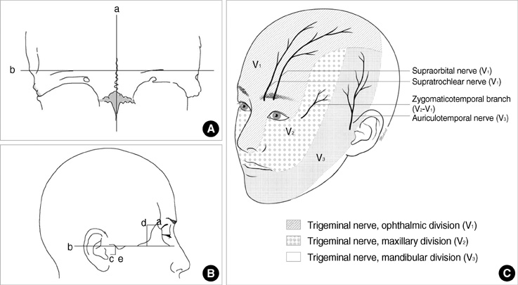
Fig. 1 Schematic drawings of the reference points and bony landmarks. (A) Reference points of the supraorbital and supratrochlear nerves. a, midline; b, supraorbital upper margin. (B) Reference points of the zygomaticotemporal and auriculotemporal nerves. a, frontozygomatic suture; b, upper margin of the zygomatic arch; c, lamina trigi; d, the point from which the zygomaticotemporal nerve exits from the deep temporalis fascia; e, the auriculotemporal nerve at its exit point; line a-d, the distance from the zygomaticofrontal suture to the zygomaticotemporal nerve; line b-d, the distance from the zygomaticotemporal nerve to the zygomatic arch upper margin; line b-e, the distance from the upper margin of the zygomatic arch to the auriculotemporal nerve at its exit point; line c-e, the distance from the auriculotemporal nerve at its exit point lamina trigi. (C) Schematic drawings of the relationship between the skin area innervated by each nerve and the subcutaneous nerve location.
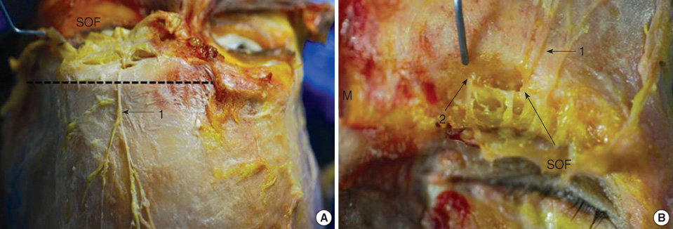
Fig. 2 Photographs of the supraorbital and supratrochlear nerve (A, B). Cadaveric photographs showing the supraorbital nerve (1) and supratrochlear nerve (2). Supraorbital rim upper margin was drawn as a dotted line. M, midline; SOF, supraorbital foramen.

Fig. 3 Photograph of zygomaticotemporal nerve and schematic drawings of the zygomaticotemporal nerve branches. (A) Cadaveric photographs showing the zygomaticotemporal nerve (1) and the frontozygomatic suture (2), (B) The nerve is classified into three types depending on the branch pattern. T, temporalis muscle.
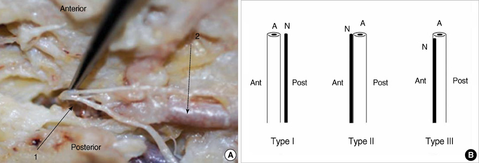
Fig. 4 Photograph of auriculotemporal nerve and schematic drawings of the relationship between the auriculotemporal nerve and the superficial temporal artery. (A) Cadaveric photographs showing the auriculotemporal nerve (1) and superficial temporal artery (2), (B) Schematic drawing of the relationship between the auriculotemporal nerve and the superficial temporal artery. Type I, The nerve runs posterior to the artery; Type II, The nerve runs anterior to artery; Type III, The nerve runs superficially parallel to the artery. A, artery; N, nerve, Ant, anterior, Post, posterior.
Reference
-
1. De Benedittis G, Lorenzetti A, Migliore M, Spagnoli D, Tiberio F, Villani RM. Postoperative pain in neurosurgery: a pilot study in brain surgery. Neurosurgery. 1996. 38:466–469.
Article2. Gee JR, Ishaq Y, Vijayan N. Postcraniotomy headache. Headache. 2003. 43:276–278.
Article3. Rocha-Filho PA, Gherpelli JL, de Siqueira JT, Rabello GD. Post-craniotomy headache: characteristics, behaviour and effect on quality of life in patients operated for treatment of supratentorial intracranial aneurysms. Cephalalgia. 2008. 28:41–48.
Article4. Guyuron B, Kriegler JS, Davis J, Amini SB. Comprehensive surgical treatment of migraine headaches. Plast Reconstr Surg. 2005. 115:1–9.5. Guyuron B, Varghai A, Michelow BJ, Thomas T, Davis J. Corrugator supercilii muscle resection and migraine headaches. Plast Reconstr Surg. 2000. 106:429–434.
Article6. Janis JE, Ghavami A, Lemmon JA, Leedy JE, Guyuron B. The anatomy of the corrugator supercilii muscle: part II. Supraorbital nerve branching patterns. Plast Reconstr Surg. 2008. 121:233–240.
Article7. Andersen NB, Bovim G, Sjaastad O. The frontotemporal peripheral nerves. Topographic variations of the supraorbital, supratrochlear and auriculotemporal nerves and their possible clinical significance. Surg Radiol Anat. 2001. 23:97–104.
Article8. Ashkenazi A, Levin M. Three common neuralgias. How to manage trigeminal, occipital, and postherpetic pain. Postgrad Med. 2004. 116:16–18. 21–24. 31–32. passim.9. Ward JB. Greater occipital nerve block. Semin Neurol. 2003. 23:59–62.
Article10. Binder WJ, Brin MF, Blitzer A, Pogoda JM. Botulinum toxin type A (BOTOX) for treatment of migraine. Dis Mon. 2002. 48:323–335.
Article11. Mathew NT, Kaup AO. The use of Botulinum toxin type A in headache treatment. Curr Treat Options Neurol. 2002. 4:365–373.
Article12. Nguyen A, Girard F, Boudreault D, Fugere F, Ruel M, Moumdjian R, Bouthilier A, Caron JL, Bojanowski MW, Girard DC. Scalp nerve blocks decrease the severity of pain after craniotomy. Anesth Analg. 2001. 93:1272–1276.
Article13. Costello TG, Cormack JR. Anaesthesia for awake craniotomy: a modern approach. J Clin Neurosci. 2004. 11:16–19.
Article14. Lavin PJ, Workman R. Cushing syndrome induced by serial occipital nerve blocks containing corticosteroids. Headache. 2001. 41:902–904.
Article15. Ohmura S, Kawada M, Ohta T, Yamamoto K, Kobayashi T. Systemic toxicity and resuscitation in bupivacaine-, levobupivacaine-, or ropivacaine-infused rats. Anesth Analg. 2001. 93:743–748.
Article16. Beer GM, Putz R, Mager K, Schumacher M, Keil W. Variations of the frontal exit of the supraorbital nerve: an anatomic study. Plast Reconstr Surg. 1998. 102:334–341.
Article17. Cuzalina AL, Holmes JD. A simple and reliable landmark for identification of the supraorbital nerve in surgery of the forehead: an in vivo anatomical study. J Oral Maxillofac Surg. 2005. 63:25–27.
Article18. Cheng AC, Yuen HK, Lucas PW, Lam DS, So KF. Characterization and localization of the supraorbital and frontal exits of the supraorbital nerve in Chinese: an anatomic study. Ophthal Plast Reconstr Surg. 2006. 22:209–213.
Article19. Saylam C, Ozer MA, Ozek C, Gurler T. Anatomical variations of the frontal and supraorbital transcranial passages. J Craniofac Surg. 2003. 14:10–12.
Article20. Anil A, Peker T, Turgut HB, Gulekon IN, Liman F. Variations in the anatomy of the inferior alveolar nerve. Br J Oral Maxillofac Surg. 2003. 41:236–239.21. Agthong S, Huanmanop T, Chentanez V. Anatomical variations of the supraorbital, infraorbital, and mental foramina related to gender and side. J Oral Maxillofac Surg. 2005. 63:800–804.
Article22. Chung MS, Kim HJ, Kang HS, Chung IH. Locational relationship of the supraorbital notch or foramen and infraorbital and mental foramina in Koreans. Acta Anat (Basel). 1995. 154:162–166.
Article23. Gumusburun E, Katkici U, Erdil H, Sevim A, Gulec E. Variations of supraorbital traits. Morphologie. 2002. 86:19–22.24. Totonchi A, Pashmini N, Guyuron B. The zygomaticotemporal branch of the trigeminal nerve: an anatomical study. Plast Reconstr Surg. 2005. 115:273–277.25. Hwang K, Suh MS, Lee SI, Chung IH. Zygomaticotemporal nerve passage in the orbit and temporal area. J Craniofac Surg. 2004. 15:209–214.
Article26. De Benedittis G. Auriculotemporal syndrome (Frey's syndrome) presenting as tic douloureux. Report of two cases. J Neurosurg. 1990. 72:955–958.27. Gulekon N, Anil A, Poyraz A, Peker T, Turgut HB, Karakose M. Variations in the anatomy of the auriculotemporal nerve. Clin Anat. 2005. 18:15–22.
- Full Text Links
-
- Actions
-
Cited
- CITED
-
- Close
- Share
-
- Similar articles
-
- Uncommon configuration of intercostobrachial nerves, lateral roots, and absent medial cutaneous nerve of arm in a cadaveric study
- Four Cases of Cutaneous Metastasis to the Scalp from Internal Malignancies
- A Case of Metastatic Adenocarcinoma on the Scalp from the Rectum
- Anatomical Investigation of Sural Nerve and Its Contributing Nerves
- A Case of Cutaneous Metastasis of Cholangiocarcinoma-like Ulcerative Nodule on the Scalp
