Korean J Gastroenterol.
2011 May;57(5):323-326. 10.4166/kjg.2011.57.5.323.
A Case of a Retroperitoneal Schwannoma Presenting as Hypermetabolic Mass in PET-CT
- Affiliations
-
- 1Department of Internal Medicine, Chungnam National University College of Medicine, Daejeon, Korea. mhs1357@hanmail.net
- KMID: 1792791
- DOI: http://doi.org/10.4166/kjg.2011.57.5.323
Abstract
- Schwannoma is a benign neoplasm of the Schwann cells of the neural sheath. Most schwannomas occur in the head and neck, and extremities and rarely in the retroperitoneal space. The differentiation of a schwannoma from other malignant tumor or benign tumor is very difficult on a preoperative examination with ultrasonography, computed tomography or magnetic resonance imaging. Furthermore, the lesion with increased fluorodeoxyglucose uptake in PET-CT cannot exclude malignant tumor. Therefore, this lesion needs surgical excision and a histological examination with immunohistochemical staining. We report a case of schwannoma occuring in the retroperitoneal space that incidentally discovered by PET-CT for health-check up. Pathologic confirmation by laparoscopic excision was done.
Keyword
MeSH Terms
Figure
-

Fig. 1. PET-CT finding. On left pelvic area, hypermetabolic mass (max SUV 3.7) (arrow) with increased FDG uptake was seen.
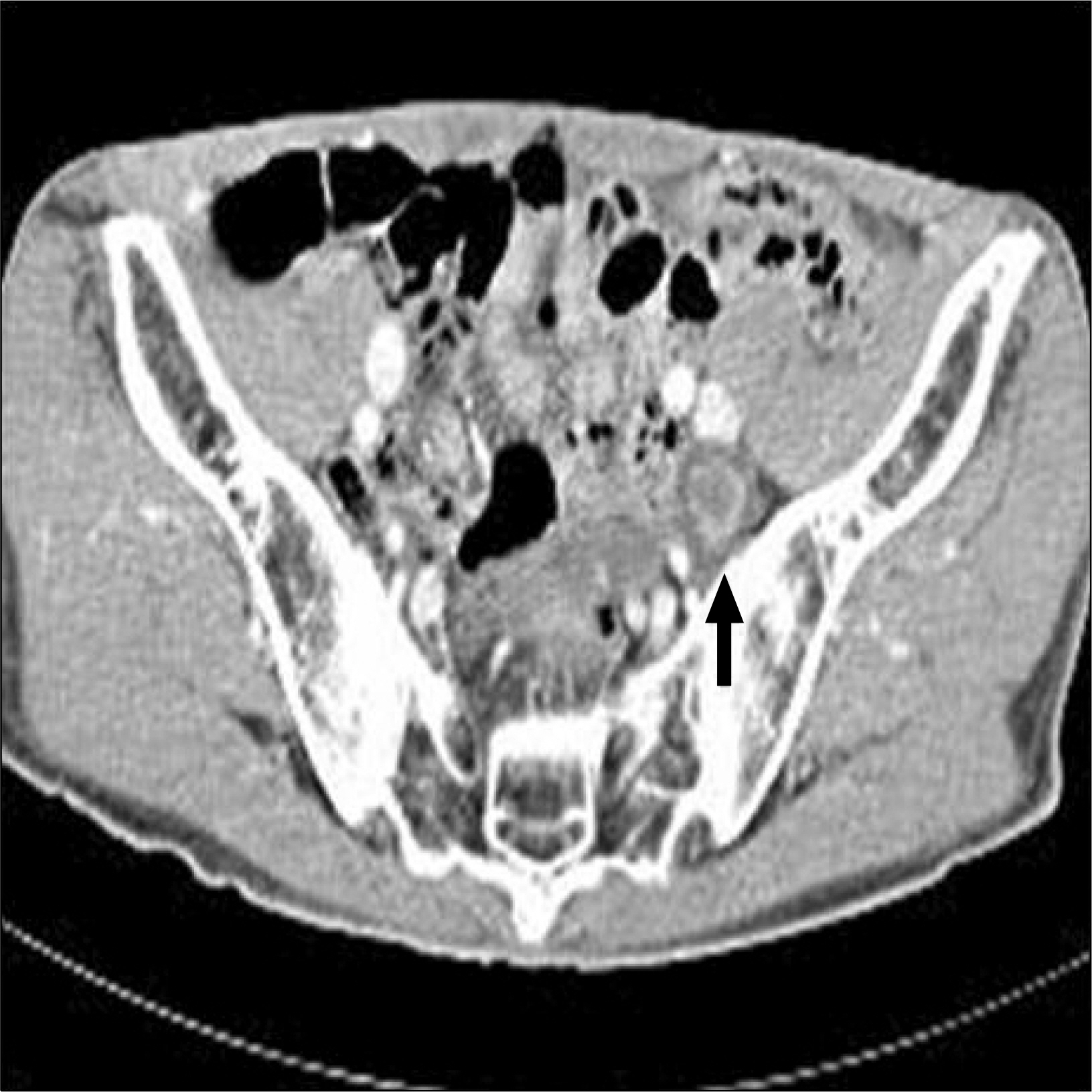
Fig. 2. Abdominal CT finding (enhanced image). Focal target-like mass (about 3×2 cm) (arrow) in adjacent to left pelvic bone was seen.

Fig. 3. Microscopic finding. It showed bundles of compact spindle cells which were surrounded by loose myxoid areas (H&E stain, ×100).
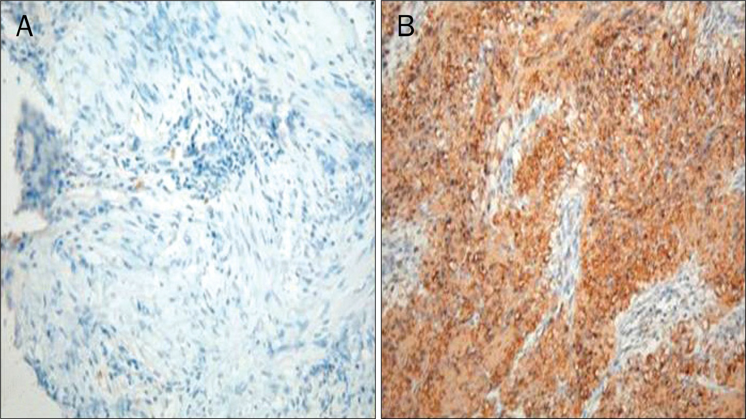
Fig. 4. Immunohistochemical staining.(A) It showed tumor cell negative for c-kit (c-kit stain, ×200). (B) Immunohistochemical stain for S-100 protein showed that tumor cells were positively statined (brown color) (S-100 protein stain, ×200).
Reference
-
References
1. Stefansson K, Wollmann R, Jerkovic M. S-100 protein in soft-tissue tumors derived from Schwann cells and melanocytes. Am J Pathol. 1982; 106:261–268.2. Thurnher D, Quint C, Pammer J, Schima W, Knerer B, Denk DM. Dysphagia due to a large schwannoma of the oropharynx: case report and review of the literature. Arch Otolaryngol Head Neck Surg. 2002; 128:850–852.3. Sharma SK, Koleski FC, Husain AN, Albala DM, Turk TM. Retroperitoneal schwannoma mimicking an adrenal lesion. World J Urol. 2002; 20:232–233.
Article4. Das Gupta TK, Brasfield RD, Strong EW, Hajdu SI. Benign solitary Schwannomas (neurilemomas). Cancer. 1969; 24:355–366.
Article5. Hide IG, Baudouin CJ, Murray SA, Malcolm AJ. Giant ancient schwannoma of the pelvis. Skeletal Radiol. 2000; 29:538–542.
Article6. Schindler OS, Dixon JH, Case P. Retroperitoneal giant schwannomas: report on two cases and review of the literature. J Orthop Surg (Hong Kong). 2002; 10:77–84.
Article7. Lee TY, Park JS, Sung YR, et al. A case report of neurilemmoma of the chest wall. Tuberc Respir Dis. 1997; 44:649–654.
Article8. Singh V, Kapoor R. Atypical presentations of benign retroperitoneal schwannoma: report of three cases with review of literature. Int Urol Nephrol. 2005; 37:547–549.
Article9. Funamizu N, Sasaki A, Matsumoto T, Inomata M, Shiraishi N, Kitano S. Laparoscopic resection of a retroperitoneal schwannoma behind the lesser omental sac. Surg Laparosc Endosc Percutan Tech. 2004; 14:175–177.
Article10. Kinoshita T, Naganuma H, Ishii K, Itoh H. CT features of retroperitoneal neurolemmoma. Eur J Radiol. 1998; 27:67–71.11. Li Q, Gao C, Juzi JT, Hao X. Analysis of 82 cases of retroperitoneal schwannoma. ANZ J Surg. 2007; 77:237–240.
Article12. Kawahara E, Oda Y, Ooi A, Katsuda S, Nakanishi I, Umeda S. Expression of glial fibrillary acidic protein (GFAP) in peripheral nerve sheath tumors. A comparative study of immunoreactivity of GFAP, vimentin, S-100 protein, and neurofilament in 38 schwannomas and 18 neurofibromas. Am J Surg Pathol. 1988; 12:115–120.13. Tokunaga T, Takeda S, Sumimura J, Maeda H. Esophageal schwannoma: report of a case. Surg Today. 2007; 37:500–502.
Article14. Beaulieu S, Rubin B, Djang D, Conrad E, Turcotte E, Eary JF. Positron emission tomography of schwannomas: emphasizing its potential in preoperative planning. AJR Am J Roentgenol. 2004; 182:971–974.
Article15. Shah N, Sibtain A, Saunders MI, Townsend E, Wong WL. High FDG uptake in a schwannoma: a PET study. J Comput Assist Tomogr. 2000; 24:55–56.16. Girgin C, Ozkan U, Sezer A, Tugyan N. Large pelvic schwannoma causing bilateral hydronephrosis. Int J Urol. 2003; 10:616–618.
Article
- Full Text Links
-
- Actions
-
Cited
- CITED
-
- Close
- Share
-
- Similar articles
-
- Benign Schwannoma Mimicking Metastatic Lesion on F-18 FDG PET/CT in Differentiated Thyroid Cancer
- Ovarian Mass Mimicking Malignancy: A Case Report
- A Case of Pelvic Retroperitoneal Schwannoma : Preoperatively Suspected of Malignant Adnexal Tumor
- Hypermetabolic Axillary Mass on 18F FDG PET/CT: Breast Cancer Arising from Accessory Breast Tissue
- A Case of Malignant Schwannoma in the Sacrum
