A Case of Ileal Duplication Cyst Lined by Ciliated Columnar and Squamous Epithelium
- Affiliations
-
- 1Department of Internal Medicine, Wonkwang University College of Medicine, Iksan, Korea.
- 2Department of Surgery, Wonkwang University College of Medicine, Iksan, Korea.
- 3Department of Pathology, Wonkwang University College of Medicine, Iksan, Korea. kjyun@wonkwang.ac.kr
- 4Digestive Disease Research Institute, Wonkwang University College of Medicine, Iksan, Korea.
- KMID: 1792765
- DOI: http://doi.org/10.4166/kjg.2009.54.1.42
Abstract
- Duplication is a rare congenital abnormality and may occur in any region of the gastrointestinal tract. A 19-year-old woman was admitted due to lower abdominal pain. Abdomino-pelvic CT scan showed a cystic mass interpreted as mesenteric cyst or duplication cyst. On the operation finding, it seemed to be arised from mesentery but attached to the ileum. Microscopically, the cystic wall was lined by non-keratinizing squamous, ciliated pseudostratified columnar epithelium, and ectopic gastric mucosa with two distinct muscular layers and a serosa. We report the first case of ileal duplication cyst lined by squamous and ciliated columnar epithelium in Korea.
Keyword
MeSH Terms
Figure
-
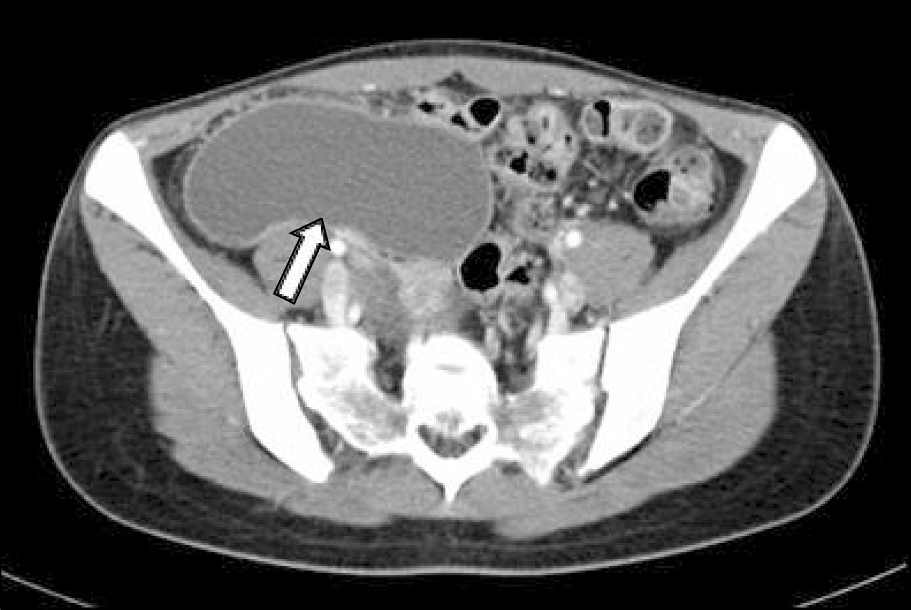
Fig. 1. Abdominopelvic CT scan showed a 8×6 cm sized, non-enhancing, thin walled, fluid-filled, cystic lesion (white arrow) on the right lower abdomen.
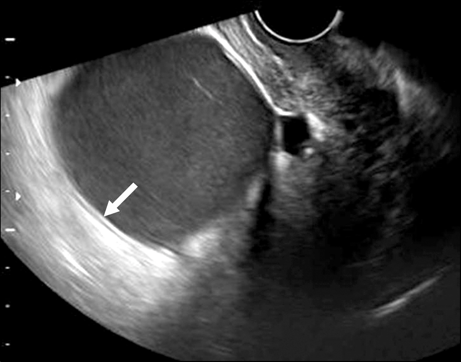
Fig. 2. Abdominal sonography showed the hypoechoic, thin walled, uniloculated cyst with inner hyperechoic mucosal and outer hypoechoic muscular layers (white arrow), consistent with a duplication.
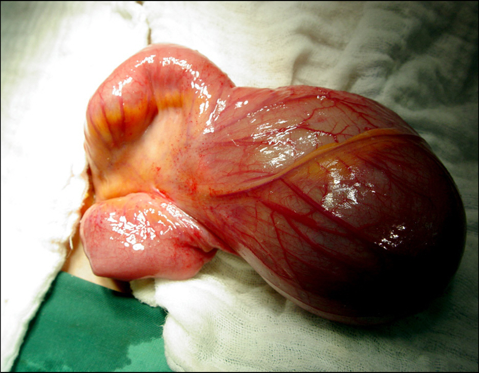
Fig. 3. Operation finding. A huge round mass was broadly based on the mesenteric side of the ileum.
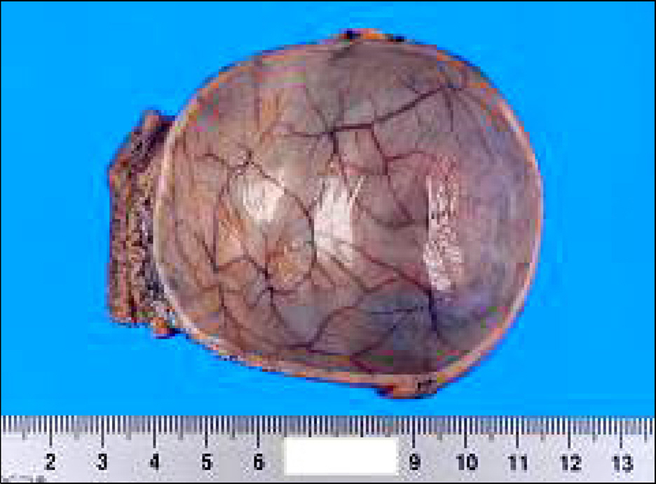
Fig. 4. Gross finding of the cross sectioned specimen showed that the cyst was uniloculated, dark serous fluid-filled and attached to the ileum without solid portion or irregular wall thickening.
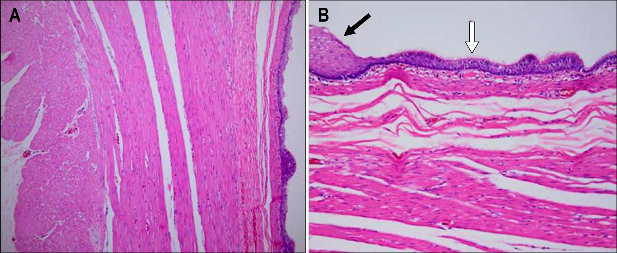
Fig. 5. Microscopic findings. (A) Two distinct muscular layers with myenteric plexus and a serosal layer attached to the small bowel wall were noted (H&E, ×100). (B) The cyst was lined by ciliated pseudostratified columnar cell (white arrow), stratified squamous cells (black arrow), and columnar cells (H&E, ×200).
Reference
-
1. Murakami S, Isozaki H, Shou T, Sakai K, Toyota H. Foregut duplication cyst of the stomach with pseudostratified columnar ciliated epithelium. Pathol Int. 2008; 58:187–190.
Article2. Porter EL. Pathology of the fetus and newborn. 2nd ed.Chicago: Year Book Medical Publishers;1961.3. Simsek A, Zeybek N, Yagci G, et al. Enteric and rectal duplications and duplication cysts in the adult. ANZ J Surg. 2005; 75:174–176.
Article4. Macpherson RI. Gastrointestinal tract duplications: clinical, pathologic, etiologic, and radiologic considerations. Radio-graphics. 1993; 13:1063–1080.
Article5. Cavar S, Bogovic M, Luetic T, Antabak A, Batinica S. Intestinal duplications - experience in 6 cases. Eur Surg Res. 2006; 38:329–332.
Article6. Teklali Y, Kaddouri N, Barahioui M. Gastrointestinal system duplications in children (19 cases). Arch Pediatr. 2002; 9:903–906.7. Kuo HC, Lee HC, Shin CH, Sheu JC, Chang PY, Wang NL. Clinical spectrum of alimentary tract duplication in children. Acta Paediatr Taiwan. 2004; 45:85–88.8. Ildstad ST, Tollerud DJ, Weiss RG, Ryan DP, McGowan MA, Martin LW. Duplications of the alimentary tract. Clinical characteristics, preferred treatment, and associated malformations. Ann Surg. 1988; 208:184–189.9. Otter MI, Marks CG, Cook MG. An unusual presentation of intestinal duplication with a literature review. Dig Dis Sci. 1996; 41:627–629.
Article10. Adair HM, Trowell JE. Squamous cell carcinoma arising in a duplication of the small bowel. J Pathol. 1981; 133:25–31.
Article11. Schiller AL, Schantz A. A cecal enterogenous cyst lined by ciliated epithelium: report of a case. Am J Clin Pathol. 1970; 53:418–422.
Article12. Dias AR, Lopes RI, do Couto RC, Bonafe WW, D'Angelo L, Salvestro ML. Ileal duplication causing recurrent intussu-sception. J Surg Educ. 2007; 64:51–53.
Article13. Gorsler C, Schier F, Danzer E. Ciliated epithelium in a midg-ut enteric duplication: a case report. Eur J Pediatr Surg. 2001; 11:136–138.
Article14. Killpack WS. Duplication of the ileum. Arch Dis Child. 1953; 28:72–75.15. Bower RJ, Sieber WK, Kiesewetter WB. Alimentary tract duplications in children. Ann Surg. 1978; 188:669–674.
Article16. Orr MM, Edwards AJ. Neoplastic change in duplications of the alimentary tract. Br J Surg. 1975; 62:269–274.
Article17. Tew K, Soans BK, Millar EA. Adenocarcinoma in an ileal duplication cyst: ultrasound and computed tomography findings. Australas Radiol. 2000; 44:228–231.
Article18. Bidwell JK, Nelson A. Prenatal ultrasonic diagnosis of congenital duplication of the stomach. J Ultrasound Med. 1986; 5:589–591.
Article19. Lee SS, Kim YH, Kang TW, et al. Recurrent gastrointestinal hemorrhage from a jejunal duplication cyst in an adult. Korean J Med. 2001; 61:264–269.20. Stringer MD, Spitz L, Abel R, et al. Management of alimentary tract duplication in children. Br J Surg. 1995; 82:74–78.
Article
