Korean J Gastroenterol.
2009 Jul;54(1):1-4. 10.4166/kjg.2009.54.1.1.
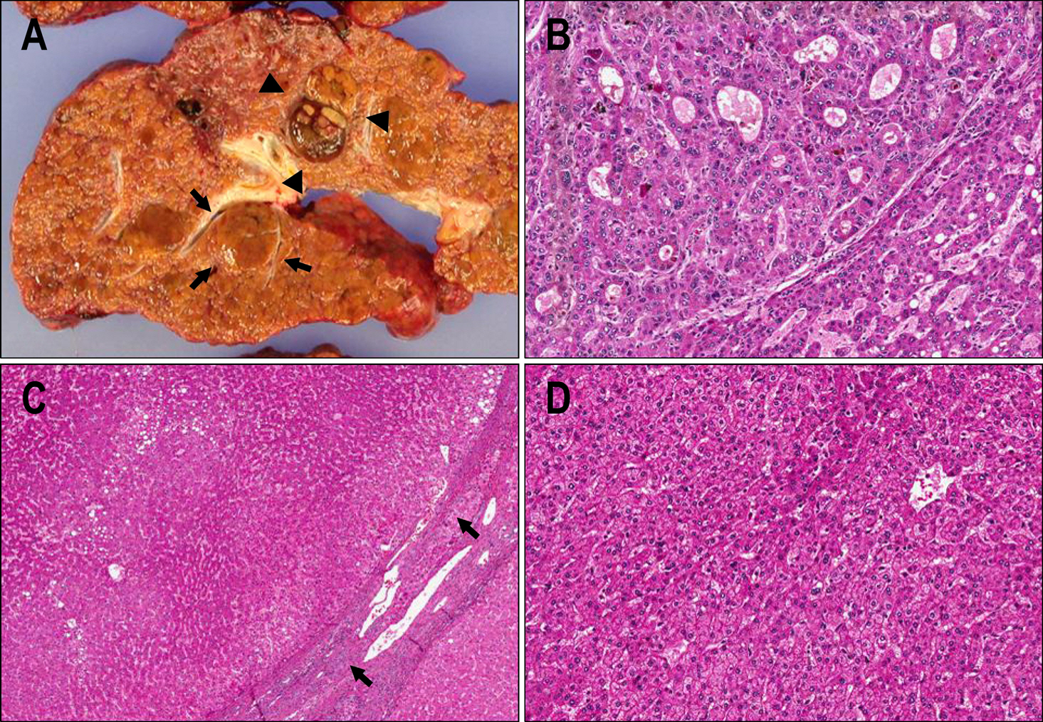
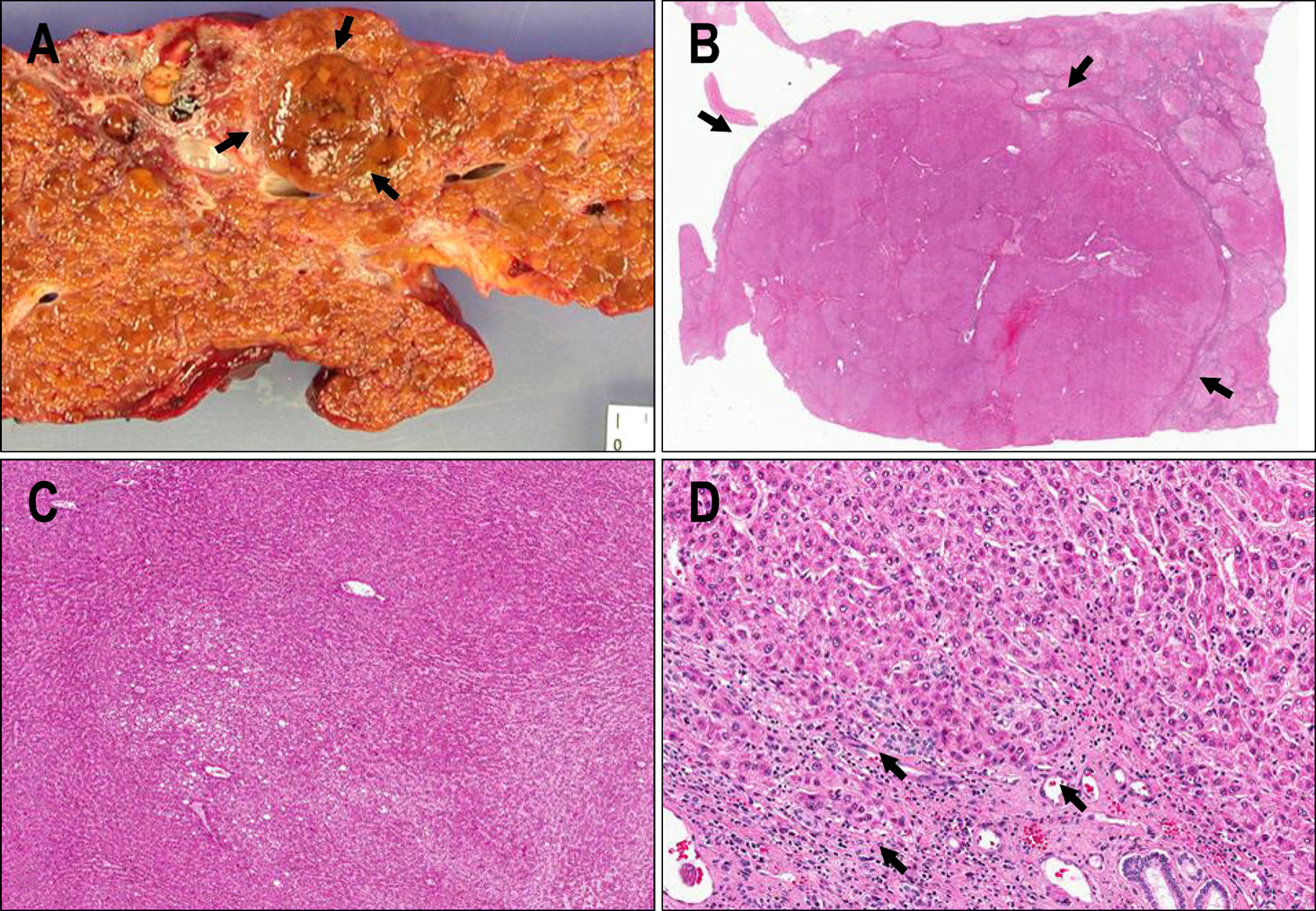
Multicentric Hepatocellular Carcinomas and Multiple Dysplastic Nodules: Spectrum of Nodular Lesions in Hepatitis B-viral Cirrhotic Liver
- Affiliations
-
- 1Department of Pathology, Ajou University College of Medicine, Suwon, Korea.
- 2Department of Pathology, Yonsei University College of Medicine and Yonsei Liver Cancer Special Clinic, Seoul, Korea. young0608@yuhs.ac
- KMID: 1792759
- DOI: http://doi.org/10.4166/kjg.2009.54.1.1
Abstract
- No abstract available.
MeSH Terms
Figure
Reference
-
1. The International Consensus Group for Hepatocellular Neoplasia. Pathologic diagnosis of early hepatocellular carcinoma: a report of the international consensus group for hepatocellular neoplasia. Hepatology. 2009; 49:658–664.2. Hytiroglou P, Park YN, Krinsky G, Theise ND. Hepatic pre-cancerous lesions and small hepatocellular carcinoma. Gastroenterol Clin N Am. 2007; 36:867–887.
Article3. Kojiro M, Roskams T. Early hepatocellular carcinoma and dysplastic nodules. Sem Liv Dis. 2005; 25:133–142.
Article4. Theise ND, Park YN, Kojiro M. Dysplastic nodules and hepatocarcinogenesis. Clin Liver Dis. 2002; 6:497–512.
Article5. Terminology of nodular hepatocellular lesions. International working party. Hepatology. 1995; 22:983–993.6. Theise N, Park C, Kim YB, Chae KJ, Park YN. Apoptosis and proliferation in hepatocarcinogenesis related to cirrhosis. Cancer. 2001; 92:2733–2738.
Article7. Theise ND, Thung SN, Cubukcu O, Fernandez GJ, Yang CP, Park YN. Neoangiogenesis and sinusoidal "capillarization" in dysplastic nodules of the liver. Am J Surg Pathol. 1998; 22:656–662.8. Park YN, Kojiro M, Di Tommaso L, et al. Ductular reaction is helpful in defining early stromal invasion, small hepatocellular carcinomas, and dysplastic nodules. Cancer. 2007; 109:915–923.
Article9. Di Tommaso L, Destro A, Seok JY, et al. The application of markers (HSP70 GPC3 and GS) in liver biopsies is useful for detection of hepatocellular carcinoma. M J Hepatol. 2009; 50:746–754.
Article
- Full Text Links
- Actions
-
Cited
- CITED
-
- Close
- Share
- Similar articles
-
- Pathology of Hepatocellular Carcinoma: Recent Update
- Hepatocarcinogenesis in liver cirrhosis: imaging diagnosis
- Focal lesions in cirrhotic liver: comparing MR imaging during arterial portography with Gd-enhanced dynamic MR imaging
- Focal nodular hyperplasia-like nodules in a young man with congenital liver cirrhosis
- A case of hypervascular hyperplastic nodules mimicking hepatocellular carcinoma in alcoholic liver cirrhosis


