Comparison of Injection of Intravitreal Drugs with Standard Care in Macular Edema Secondary to Branch Retinal Vein Occlusion
- Affiliations
-
- 1HanGil Eye Hospital, Incheon, Korea. Jhsohn19@hanafos.com
- KMID: 1792090
- DOI: http://doi.org/10.3341/kjo.2014.28.1.19
Abstract
- PURPOSE
To compare the long-term efficacy and safety of intravitreal triamcinolon with or without rescue laser therapy (intravitreal triamcinolone injection [IVTA] group), bevacizumab with or without rescue laser treatment (intravitreal bevacizumab injection [IVB] group), or a combination of both with or without rescue laser therapy (IVTA + IVB group), with standard care for patients with macular edema secondary to branch retinal vein occlusion (BRVO).
METHODS
We reviewed the medical records of 151 patients treated with intravitreal injection with or without rescue laser for treatment of macular edema caused by BRVO, and who were followed up at 1, 3, 6, 12, and 24 months. During the observation period, rescue grid laser or repeated intravitreal injection with initial drug was performed if recurrence of macular edema was confirmed. Visual acuity, change in visual acuity, and intraocular pressure were compared in each phase.
RESULTS
Totals of 16%, 5.6%, and 0% of participants in the three groups showed significant visual loss of more than three lines of the Snellen chart at last follow-up. The IVTA group was the least effective treatment modality, with statistical significance. The development rates of elevated intraocular pressure were similar among the groups.
CONCLUSIONS
Although IVTA yielded effects similar to those of standard grid photocoagulation based on the Standard Care vs Corticosteroid for Retinal Vein Occlusion study, IVB or IVTA + IVB with or without rescue laser treatment resulted in improvement in visual acuity at 24 months after the start of treatment and was associated with few serious adverse side effects. Thus, these approaches could be useful for treating macular edema arising secondary to BRVO.
MeSH Terms
-
Angiogenesis Inhibitors/administration & dosage
Antibodies, Monoclonal, Humanized/*administration & dosage
Female
Follow-Up Studies
Glucocorticoids/administration & dosage
Humans
Intravitreal Injections
Laser Therapy/*methods
Macular Edema/diagnosis/etiology/*therapy
Male
Middle Aged
Recurrence
Retinal Vein Occlusion/*complications/diagnosis/therapy
Retrospective Studies
Tomography, Optical Coherence
Treatment Outcome
Triamcinolone Acetonide/*administration & dosage
Vascular Endothelial Growth Factor A/*antagonists & inhibitors
Visual Acuity
Angiogenesis Inhibitors
Antibodies, Monoclonal, Humanized
Glucocorticoids
Triamcinolone Acetonide
Vascular Endothelial Growth Factor A
Figure
-
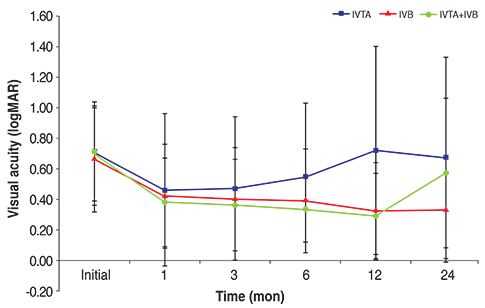
Fig. 1 Visual acuity as measured by a Snellen chart at each follow-up interval. The three groups showed no statistically significant difference in visual acuity in most of the follow-up interval except for the 12-month follow-up, at which time the intravitreal triamcinolone injection (IVTA) group showed a statistically significant decrease in visual acuity (graphed on the logarithm of the minimum angle of resolution [logMAR] scale). IVB = intravitreal bevacizumab injection.
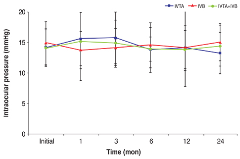
Fig. 2 Intraocular pressure at each follow-up interval. The three groups showed no statistically significant differences in intraocular pressure at any follow-up interval. IVTA = intravitreal triamcinolone injection; IVB = intravitreal bevacizumab injection.
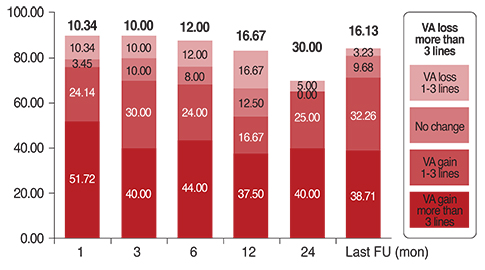
Fig. 3 Patient distribution in the intravitreal triamcinolone injection group with respect to change in visual acuity (VA) differences as measured using a Snellen chart at each follow-up (FU) interval. The levels reflect gain or loss of Snellen chart lines compared with baseline VA.
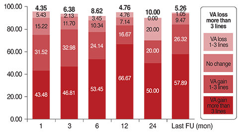
Fig. 4 Patient distribution in the intravitreal bevacizumab injection group with respect to change in visual acuity (VA) differences as measured using a Snellen chart at each follow-up (FU) interval. The levels reflect gain or loss of Snellen chart lines compared with baseline VA.
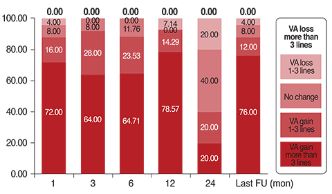
Fig. 5 Patient distribution in the intravitreal triamcinolone injection + intravitreal bevacizumab injection group with respect to change in visual acuity (VA) differences as measured using a Snellen chart at each follow-up (FU) interval. The levels reflect gain or loss of Snellen chart lines compared with baseline VA.
Cited by 3 articles
-
Combined Low Dose Bevacizumab-triamcinolone versus Bevacizumab Single Intravitreal Injection for Branch Retinal Vein Occlusion
Min Ho Shin, Hyun Ji Kang, Jin Seok Seo, In Young Chung
J Korean Ophthalmol Soc. 2018;59(7):650-656. doi: 10.3341/jkos.2018.59.7.650.Comparison of Bevacizumab and Combined Low-dose Bevacizumab and Triamcinolone in Central Retinal Vein Occlusion
Byung Jae Kim, Hyun Woong Kim, Yong Seop Han, Jong Moon Park, In Young Chung
J Korean Ophthalmol Soc. 2016;57(3):438-444. doi: 10.3341/jkos.2016.57.3.438.Natural Short-term Course of Recurrent Macular Edema Following Intravitreal Bevacizumab Therapy in Branch Retinal Vein Occlusion
Su Jin Yoo, Jae Hui Kim, Tae Gon Lee, Jong Woo Kim, Sung Won Cho, Jung Il Han
Korean J Ophthalmol. 2017;31(2):95-101. doi: 10.3341/kjo.2017.31.2.95.
Reference
-
1. Hayreh SS. Prevalent misconceptions about acute retinal vascular occlusive disorders. Prog Retin Eye Res. 2005; 24:493–519.2. Wong TY, Larsen EK, Klein R, et al. Cardiovascular risk factors for retinal vein occlusion and arteriolar emboli: the Atherosclerosis Risk in Communities & Cardiovascular Health studies. Ophthalmology. 2005; 112:540–547.3. Mitchell P, Smith W, Chang A. Prevalence and associations of retinal vein occlusion in Australia: the Blue Mountains Eye Study. Arch Ophthalmol. 1996; 114:1243–1247.4. Klein R, Klein BE, Moss SE, Meuer SM. The epidemiology of retinal vein occlusion: the Beaver Dam Eye Study. Trans Am Ophthalmol Soc. 2000; 98:133–141.5. Klein R, Moss SE, Meuer SM, Klein BE. The 15-year cumulative incidence of retinal vein occlusion: the Beaver Dam Eye Study. Arch Ophthalmol. 2008; 126:513–518.6. Cugati S, Wang JJ, Rochtchina E, Mitchell P. Ten-year incidence of retinal vein occlusion in an older population: the Blue Mountains Eye Study. Arch Ophthalmol. 2006; 124:726–732.7. The Branch Vein Occlusion Study Group. Argon laser photocoagulation for macular edema in branch vein occlusion. Am J Ophthalmol. 1984; 98:271–282.8. Chang MA, Fine HF, Bass E, et al. Patients' preferences in choosing therapy for retinal vein occlusions. Retina. 2007; 27:789–797.9. McIntosh RL, Mohamed Q, Saw SM, Wong TY. Interventions for branch retinal vein occlusion: an evidence-based systematic review. Ophthalmology. 2007; 114:835–854.10. Scott IU, Ip MS, VanVeldhuisen PC, et al. A randomized trial comparing the efficacy and safety of intravitreal triamcinolone with standard care to treat vision loss associated with macular Edema secondary to branch retinal vein occlusion: the Standard Care vs Corticosteroid for Retinal Vein Occlusion (SCORE) study report 6. Arch Ophthalmol. 2009; 127:1115–1128.11. Campochiaro PA, Heier JS, Feiner L, et al. Ranibizumab for macular edema following branch retinal vein occlusion: six-month primary end point results of a phase III study. Ophthalmology. 2010; 117:1102–1112.e1.12. Haller JA, Bandello F, Belfort R Jr, et al. Dexamethasone intravitreal implant in patients with macular edema related to branch or central retinal vein occlusion twelve-month study results. Ophthalmology. 2011; 118:2453–2460.13. Rogers SL, McIntosh RL, Lim L, et al. Natural history of branch retinal vein occlusion: an evidence-based systematic review. Ophthalmology. 2010; 117:1094–1101.e5.14. Guthoff R, Meigen T, Hennemann K, Schrader W. Comparison of bevacizumab and triamcinolone for treatment of macular edema secondary to branch retinal vein occlusion in a pair-matched analysis. Ophthalmologica. 2010; 224:319–324.15. Ehrlich R, Ciulla TA, Moss AM, Harris A. Combined treatment of intravitreal bevacizumab and intravitreal triamcinolone in patients with retinal vein occlusion: 6 months of follow-up. Graefes Arch Clin Exp Ophthalmol. 2010; 248:375–380.16. Cekic O, Cakir M, Yazici AT, et al. A comparison of three different intravitreal treatment modalities of macular edema due to branch retinal vein occlusion. Curr Eye Res. 2010; 35:925–929.
- Full Text Links
-
- Actions
-
Cited
- CITED
-
- Close
- Share
-
- Similar articles
-
- Effects of Intravitreal Bevacizumab Injection in 3 Types of Macular Edema Secondary to Branch Retinal Vein Occlusion
- The Efficacy of Intravitreal Bevacizumab in the Treatment of Macular Edema
- Short-term Outcomes of Ranibizumab Biosimilar CKD-701 Treatment in Macular Edema Secondary to Retinal Vein Occlusion
- Intravitreal Bevacizumab Injection for Macular Edema Secondary to Branch Retinal Vein Occlusion: Long-Term Results
- The Effectiveness of Intravitreal Triamcinolone Injection for the Treatment of Macular Edema
