Long-term Successful Treatment of Massive Distal Duodenal Variceal Bleeding with Balloon-occluded Retrograde Transvenous Obliteration
- Affiliations
-
- 1Department of Internal Medicine, Hanyang University Guri Hospital, Hanyang University College of Medicine, Guri, Korea. sonjh@hanyang.ac.kr
- 2Department of Radiology, Hanyang University Seoul Hospital, Hanyang University College of Medicine, Seoul, Korea.
- KMID: 1791883
- DOI: http://doi.org/10.4166/kjg.2014.63.4.248
Abstract
- Duodenal variceal bleeding in patients with portal hypertension due to cirrhosis or other causes is uncommon. We report on a case of a 55-year-old male with an ectopic variceal rupture at the distal fourth part of the duodenum who presented with massive hematochezia and shock. Shortly after achievement of hemodynamic stability, due to the limitation of an endoscopic procedure, we initially attempted to find the bleeding focus by abdominal computed tomography, which showed tortuous duodenal varices that drained into the left gonadal vein. He was treated with first-line balloon-occluded retrograde transvenous obliteration (BRTO), resulting in a favorable long-term outcome without rebleeding three years later. This case suggests that BRTO may be a first-line therapeutic option for control of ruptured duodenal varices, especially at a distal location.
MeSH Terms
Figure
-
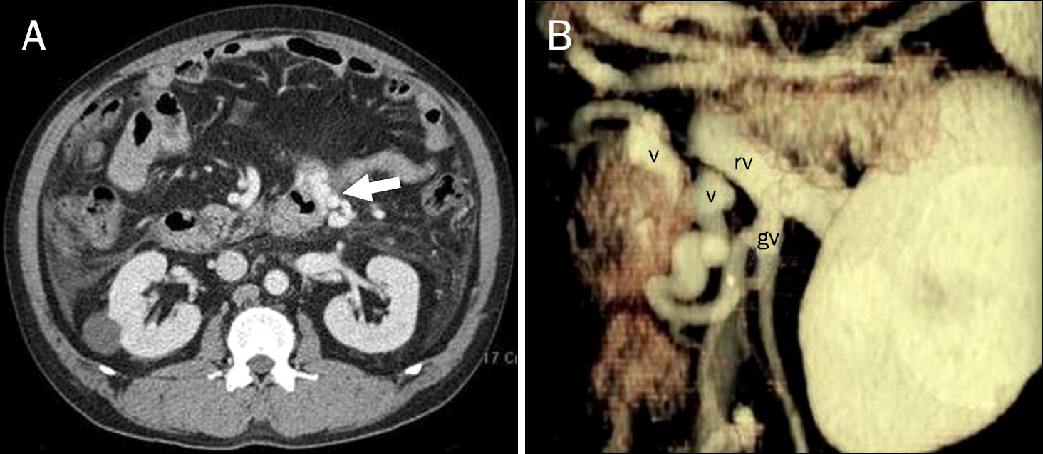
Fig. 1. (A) Axial images of a portal- phase abdominal CT scan show varices (arrow) of the fourth part of the duodenum. (B) These duodenal varices (v) drained into the left renal vein (rv) through the left gonadal vein (gv).

Fig. 2. (A) Radiograph obtained after infusion of a mixture of a sclerotic agent and lipiodol through a micro-catheter (white arrow) during balloon occlusion (black arrow) shows duodenal varices and the afferent vein (asterisks). (B) Axial image of a portal phased abdominal CT shows filling of the sclerosant within duodenal varices by the sclerosant (arrow).
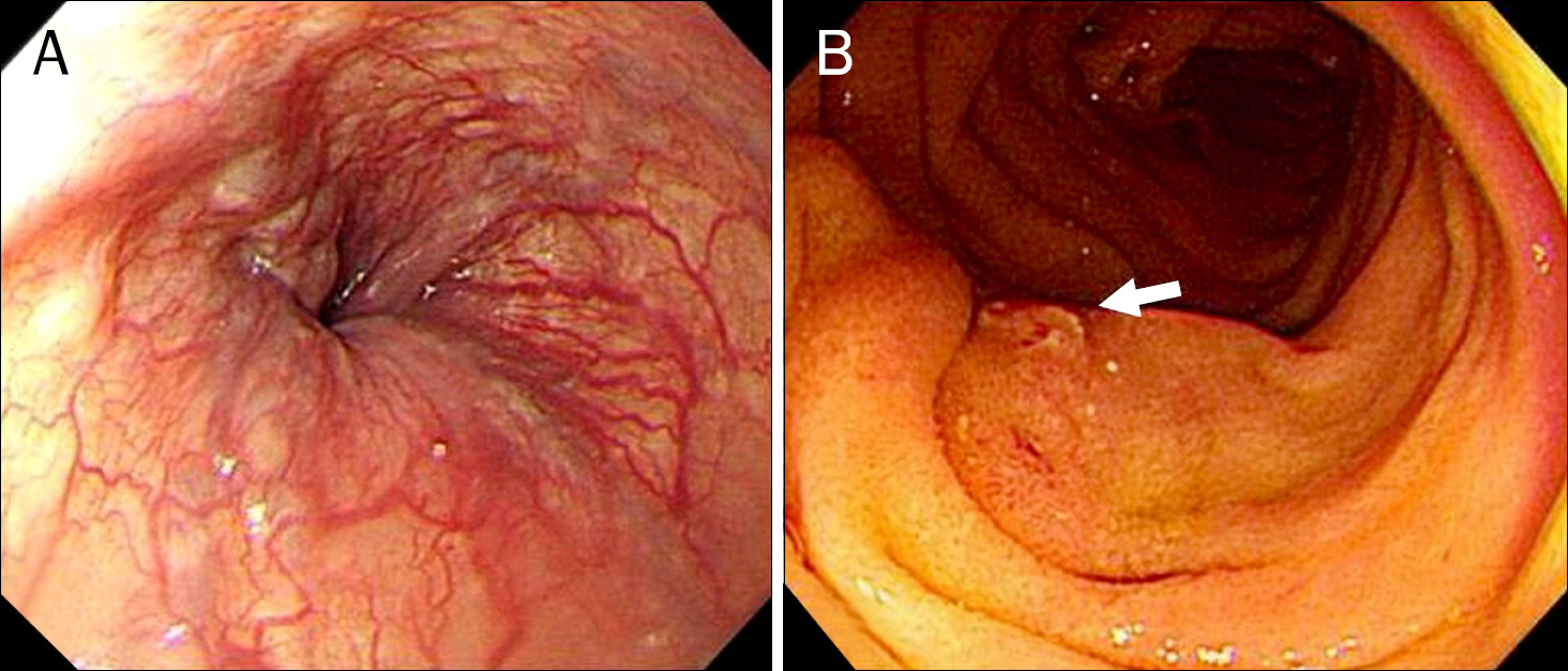
Fig. 3. (A) Two weeks after the balloon-occluded retrograde transvenous obliteration procedure, gastroduodenoscopy showed small esophageal varices at the lower esophagus. (B) Push enteroscopy showed ruptured erosion (arrow) on the varix at the fourth part of the duodenum.
Reference
-
References
1. Helmy A, Al Kahtani K, Al Fadda M. Updates in the pathogenesis, diagnosis and management of ectopic varices. Hepatol Int. 2008; 2:322–334.
Article2. Kanagawa H, Mima S, Kouyama H, Gotoh K, Uchida T, Okuda K. Treatment of gastric fundal varices by balloon-occluded retrograde transvenous obliteration. J Gastroenterol Hepatol. 1996; 11:51–58.
Article3. Ohta M, Yasumori K, Saku M, Saitsu H, Muranaka T, Yoshida K. Successful treatment of bleeding duodenal varices by balloon-occluded retrograde transvenous obliteration: a transjugular venous approach. Surgery. 1999; 126:581–583.
Article4. Tominaga K, Montani A, Kuga T, et al. Combined balloon-oc-cluded embolization for treatment of concurrent duodenal, gastric, and esophageal varices: a case report. Gastrointest Endosc. 2001; 53:665–668.
Article5. Ota K, Okazaki M, Higashihara H, et al. Combination of tran-sileocolic vein obliteration and balloon-occluded retrograde transvenous obliteration is effective for ruptured duodenal varices. J Gastroenterol. 1999; 34:694–699.
Article6. Takamura K, Miyake H, Mori H, et al. Balloon occluded retrograde transvenous obliteration and percutaneous transhepatic obliteration for ruptured duodenal varices after operation for rectal cancer with multiple liver metastasis: report of a case. J Med Invest. 2005; 52:212–217.
Article7. Zamora CA, Sugimoto K, Tsurusaki M, et al. Endovascular obliteration of bleeding duodenal varices in patients with liver cirrhosis. Eur Radiol. 2006; 16:73–79.
Article8. Tanaka O, Ohno K, Ohno T, et al. Should balloon-occluded retrograde transvenous obliteration be the first-line interventional radiologic treatment for bleeding duodenal varices? A case report and review of the literature. Acta Radiol. 2008; 49:32–36.
Article9. Norton ID, Andrews JC, Kamath PS. Management of ectopic varices. Hepatology. 1998; 28:1154–1158.
Article10. Fayad N, Nammour F, Elfant A. Endoscopic variceal ligation for bleeding duodenal varices. J Clin Gastroenterol. 2004; 38:467.
Article11. Barbish AW, Ehrinpreis MN. Successful endoscopic injection sclerotherapy of a bleeding duodenal varix. Am J Gastroenterol. 1993; 88:90–92.12. Almeida JR, Trevisan L, Guerrazzi F, et al. Bleeding duodenal varices successfully treated with TIPS. Dig Dis Sci. 2006; 51:1738–1741.
Article13. Son BK, Sohn JH, Chang MH, Park YK, Kim TY, Jeon YC. A case of successful endoscopic injection sclerotherapy with N-bu-tyl-2-cyanoacrylate for ruptured duodenal varices. Korean J Gastroenterol. 2007; 49:336–340.14. Amin R, Alexis R, Korzis J. Fatal ruptured duodenal varix: a case report and review of literature. Am J Gastroenterol. 1985; 80:13–18.15. Park SB, Lee SH, Kim JH, et al. Successful treatment of duodenal variceal bleeding by endoscopic clipping. Clin Endosc. 2013; 46:403–406.
Article16. Akazawa Y, Murata I, Yamao T, et al. Successful management of bleeding duodenal varices by endoscopic variceal ligation and balloon-occluded retrograde transvenous obliteration. Gastrointest Endosc. 2003; 58:794–797.
Article17. Hashimoto R, Sofue K, Takeuchi Y, Shibamoto K, Arai Y. Successful balloon-occluded retrograde transvenous obliteration for bleeding duodenal varices using cyanoacrylate. World J Gastroenterol. 2013; 19:951–954.
Article18. Ponec RJ, Kowdley KV. Paradoxical cerebral emboli after transjugular intrahepatic portosystemic shunt and coil embolization for treatment of duodenal varices. Am J Gastroenterol. 1997; 92:1372–1373.19. Koito K, Namieno T, Nagakawa T, Morita K. Balloon-occluded retrograde transvenous obliteration for gastric varices with gastrorenal or gastrocaval collaterals. AJR Am J Roentgenol. 1996; 167:1317–1320.
Article
- Full Text Links
-
- Actions
-
Cited
- CITED
-
- Close
- Share
-
- Similar articles
-
- Plug-Assisted Retrograde Transvenous Obliteration for the Treatment of Duodenal Variceal Bleeding - A Case Report and Literature Review
- Successful Treatment of Duodenal Variceal Bleeding with Coil-Assisted Retrograde Transvenous Obliteration: A Case Report
- Plug-Assisted Retrograde Transvenous Obliteration for the Treatment of Gastric Varix with Both Gastrorenal and Gastrocaval Shunts: A Case Report
- Balloon Occluded Retrograde Transvenous Obliteration of Bleeding Stomal Varices Using Sodium Tetradecyl Sulfate Foam: A Case Report
- Percutaneous transvenous obliteration for duodenal variceal bleeding using micro-balloon catheter and n-butyl cyanoacrylate: A case report
