Safety and Effectiveness of Successive Extracorporeal Shock Wave Lithotripsy for Pancreatolithiasis under Intravenous Bolus Pethidine Administration Alone
- Affiliations
-
- 1Department of Gastroenterology, Ulsan University Hospital, University of Ulsan College of Medicine, Ulsan, Korea.
- 2Division of Gastroenterology, Department of Internal Medicine, Asan Medical Center, Seoul, Korea. mhkim@amc.seoul.kr
- KMID: 1791880
- DOI: http://doi.org/10.4166/kjg.2014.63.4.231
Abstract
- BACKGROUND/AIMS
A retrospective analysis was performed to evaluate the safety and effectiveness of extracorporeal shock wave lithotripsy (ESWL) for pancreatolithiasis on successive days under intravenous bolus of pethidine alone.
METHODS
Ninety patients with calcified pancreatic stones (> or =5 mm) presenting with abdominal pain were selected for ESWL. ESWL was performed with an electroconductive lithotripter under fluoroscopic target systems. Fragmented calculi after ESWL were removed by endotherapy.
RESULTS
A mean of 4.2 ESWL sessions were performed for each patient, with a mean of 2,984 shocks at a mean power setting of 12.8 kV. Eighty-four (89.3%) patients underwent ESWL for three or more days in a row. Fragmentation of the stones were achieved in 83/90 (92.2%) patients. Complete clearance of the main pancreatic duct was achieved in 54/90 (60.0%) patients, and partial clearance was achieved in 27 (30.0%) patients. The mean dose of pethidine used during ESWL was 53.5+/-20.7 mg per session. As ESWL-related complications, four (4.3%) patients developed mild acute pancreatitis.
CONCLUSIONS
In case of endoscopically difficult-to-treat pancreatic duct stones, combined therapy with ESWL is an effective method, and treatment with multiple sessions of ESWL on successive days under intravenous bolus of pethidine alone is safe and well tolerated.
MeSH Terms
Figure
-
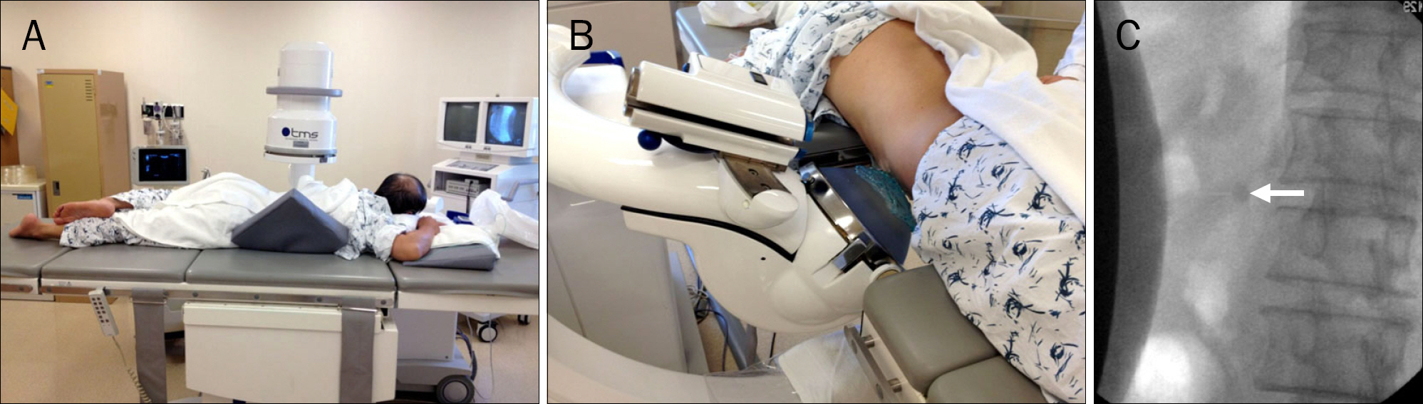
Fig. 1. (A, B) During extracorporeal shock wave lithotripsy, the patient lies on the lithotripter table in the prone position. (C) Fluoroscopy image shows a targeted stone (arrow).
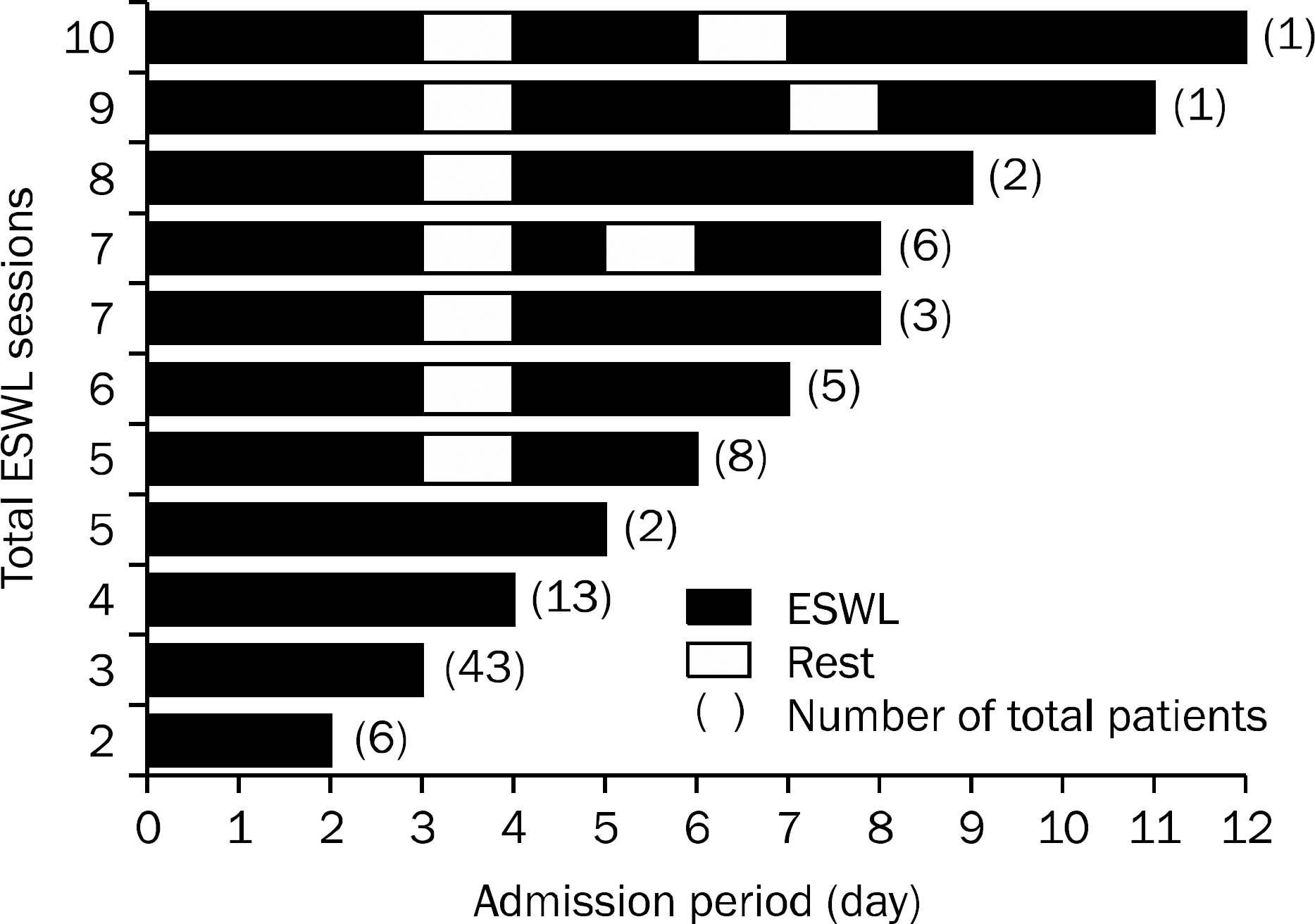
Fig. 2. Extracorporeal shock wave lithotripsy (ESWL) sessions provided during the same admission period in an individual patient.
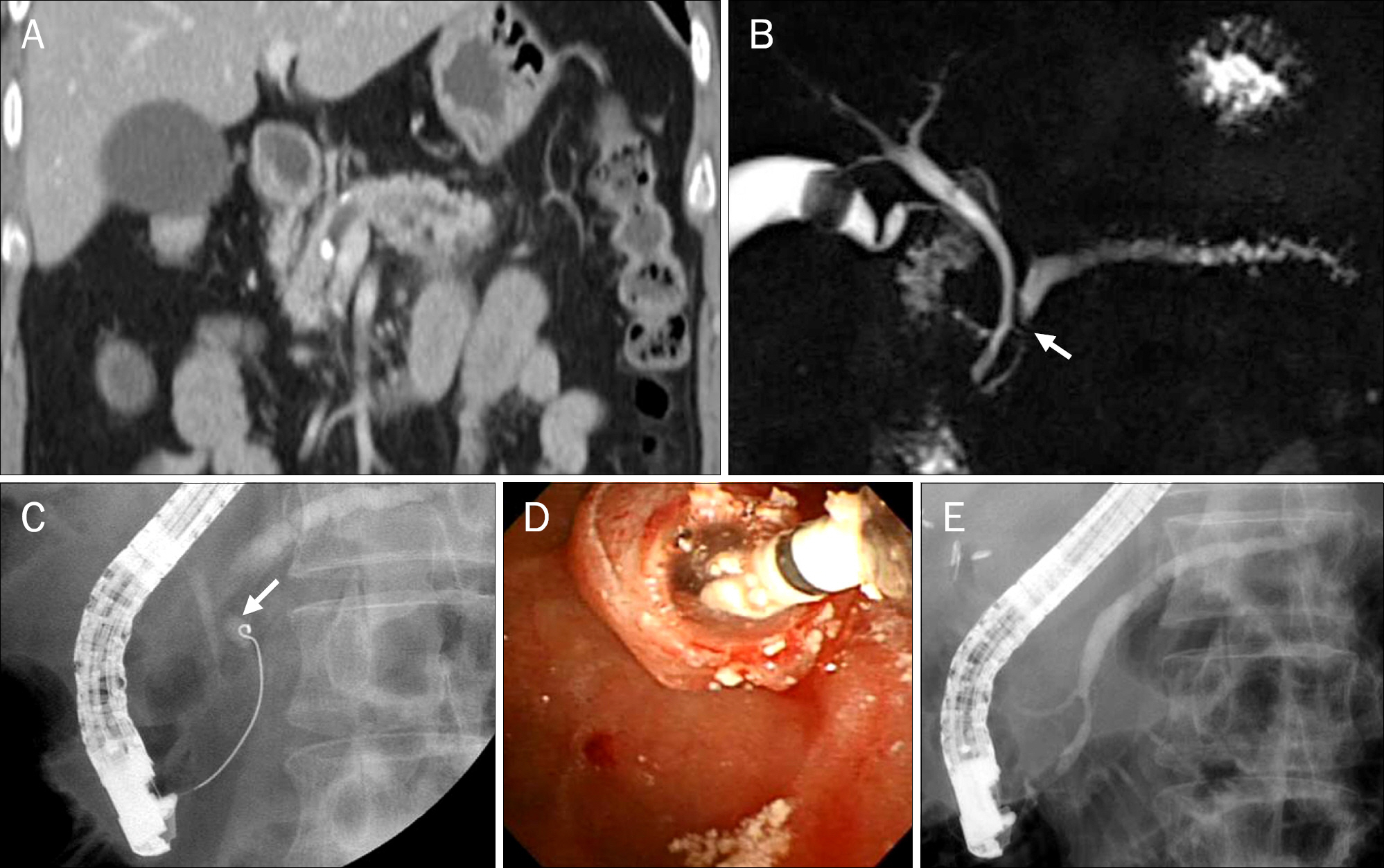
Fig. 3. (A-C) Pre-extracorporeal shock wave lithotripsy (ESWL); (A, B) Abdominal CT and MRCP show an impacted pancreatic stone (arrow) with upstream duct dilatation. (C) ERCP reveals a partially opacified main pancreatic duct with contrast injection. However, the guidewire failed to advance beyond the impacted stone (arrow) into the upstream duct. (D, E) Post-ESWL; (D) Fragmented pancreatic duct stones by ESWL were removed by balloon catheter during ERCP. (E) Follow-up pancreatogram shows no residual pancreatolithiasis in the main pancreatic duct.
Cited by 1 articles
-
First-line Treatment for Chronic Pancreatitis with Stones: Extracorporeal Shock Wave Lithotripsy?
Seok Ho Dong
Korean J Gastroenterol. 2014;63(4):199-200. doi: 10.4166/kjg.2014.63.4.199.
Reference
-
References
1. Etemad B, Whitcomb DC. Chronic pancreatitis: diagnosis, classification, and new genetic developments. Gastroenterology. 2001; 120:682–707.
Article2. Ammann RW, Muench R, Otto R, Buehler H, Freiburghaus AU, Siegenthaler W. Evolution and regression of pancreatic calcification in chronic pancreatitis. A prospective long-term study of 107 patients. Gastroenterology. 1988; 95:1018–1028.3. Bhasin DK, Singh G, Rana SS, et al. Clinical profile of idiopathic chronic pancreatitis in North India. Clin Gastroenterol Hepatol. 2009; 7:594–599.
Article4. Matthews K, Correa RJ, Gibbons RP, Weissman RM, Kozarek RA. Extracorporeal shock wave lithotripsy for obstructing pancreatic duct calculi. J Urol. 1997; 158:522–525.
Article5. Ebbehøj N, Borly L, Bülow J, Rasmussen SG, Madsen P. Evaluation of pancreatic tissue fluid pressure and pain in chronic pancreatitis. A longitudinal study. Scand J Gastroenterol. 1990; 25:462–466.
Article6. Widdison AL, Alvarez C, Karanjia ND, Reber HA. Experimental evidence of beneficial effects of ductal decompression in chronic pancreatitis. Endoscopy. 1991; 23:151–154.
Article7. Tandan M, Reddy DN, Talukdar R, et al. Longterm clinical outcomes of extracorporeal shockwave lithotripsy in painful chronic calcific pancreatitis. Gastrointest Endosc. 2013; 78:726–733.
Article8. Dumonceau JM, Delhaye M, Tringali A, et al. Endoscopic treatment of chronic pancreatitis: European Society of Gastrointestinal Endoscopy (ESGE) Clinical Guideline. Endoscopy. 2012; 44:784–800.
Article9. Inui K, Tazuma S, Yamaguchi T, et al. Treatment of pancreatic stones with extracorporeal shock wave lithotripsy: results of a multicenter survey. Pancreas. 2005; 30:26–30.10. Delhaye M, Vandermeeren A, Baize M, Cremer M. Extracorporeal shockwave lithotripsy of pancreatic calculi. Gastroenterology. 1992; 102:610–620.
Article11. Dumonceau JM, Costamagna G, Tringali A, et al. Treatment for painful calcified chronic pancreatitis: extracorporeal shock wave lithotripsy versus endoscopic treatment: a randomised controlled trial. Gut. 2007; 56:545–552.
Article12. Tandan M, Reddy DN, Santosh D, et al. Extracorporeal shock wave lithotripsy and endotherapy for pancreatic calculi-a large single center experience. Indian J Gastroenterol. 2010; 29:143–148.
Article13. McCaffery M, Pasero C. Pain: clinical manual. 2nd ed.St. Louis: Mosby;1999.14. Banks PA, Bollen TL, Dervenis C, et al. Acute Pancreatitis Classification Working Group. Classification of acute pancreatitis–2012: revision of the Atlanta classification and definitions by international consensus. Gut. 2013; 62:102–111.
Article15. Choi KS, Kim MH, Lee YS, et al. Disintegration of pancreatic duct stones with extracorporeal shockwave lithotripsy. Korean J Gastroenterol. 2005; 46:396–403.16. Pemberton RJ, Tolley DA. Comparison of a new-generation electroconductive spark lithotripter and the Dornier Compact Delta for ureteral calculi in a quaternary referral center. J Endourol. 2006; 20:732–736.
Article17. Schlosser W, Beger HG. Organ-preserving surgery in chronic pancreatitis: the duodenum-preserving pancreatic head resection. Ann Ital Chir. 2000; 71:65–70.18. Sherman S, Lehman GA, Hawes RH, et al. Pancreatic ductal stones: frequency of successful endoscopic removal and improvement in symptoms. Gastrointest Endosc. 1991; 37:511–517.
Article19. Ohara H, Hoshino M, Hayakawa T, et al. Single application extracorporeal shock wave lithotripsy is the first choice for patients with pancreatic duct stones. Am J Gastroenterol. 1996; 91:1388–1394.20. Suzuki Y, Sugiyama M, Inui K, et al. Management for pancreatolithiasis: a Japanese multicenter study. Pancreas. 2013; 42:584–588.21. Lawrence C, Siddiqi MF, Hamilton JN, et al. Chronic calcific pancreatitis: combination ERCP and extracorporeal shock wave lithotripsy for pancreatic duct stones. South Med J. 2010; 103:505–508.
Article22. Kozarek RA, Brandabur JJ, Ball TJ, et al. Clinical outcomes in patients who undergo extracorporeal shock wave lithotripsy for chronic calcific pancreatitis. Gastrointest Endosc. 2002; 56:496–500.
Article23. Johanns W, Jakobeit C, Greiner L, Janssen J. Ultrasound-guided extracorporeal shock wave lithotripsy of pancreatic ductal stones: six years' experience. Can J Gastroenterol. 1996; 10:471–475.
Article24. Sasahira N, Tada M, Isayama H, et al. Outcomes after clearance of pancreatic stones with or without pancreatic stenting. J Gastroenterol. 2007; 42:63–69.
Article25. Seven G, Schreiner MA, Ross AS, et al. Longterm outcomes associated with pancreatic extracorporeal shock wave lithotripsy for chronic calcific pancreatitis. Gastrointest Endosc. 2012; 75:997–1004.
Article
- Full Text Links
-
- Actions
-
Cited
- CITED
-
- Close
- Share
-
- Similar articles
-
- Clinical Experience of Extracorporeal Shock Wave Lithotripsy for Urinary Calculi
- Effects of Extracorporeal Shock Wave Lithortripsy Experimentally Induced Cholelithiasis and Organs in the Dog
- Extracorporeal shock wave lithotripsy of lower caliceal stone
- The effect of double-J stent in extracorporeal shock wave lithotripsy monotherapy of staghorn calculi
- Sedative - Analgesic Effect with Diazepam - Fentanyl for Extracorporeal Shock Wave Lithotripsy
