Blood Res.
2015 Mar;50(1):61-62. 10.5045/br.2015.50.1.61.
Diffuse large B cell lymphoma with high M protein: an unusual finding
- Affiliations
-
- 1Department of Pathology and Laboratory Medicine, Medanta-The Medicity, Gurgaon, India. sachdev05@gmail.com
- KMID: 1787917
- DOI: http://doi.org/10.5045/br.2015.50.1.61
Abstract
- No abstract available.
MeSH Terms
Figure
-
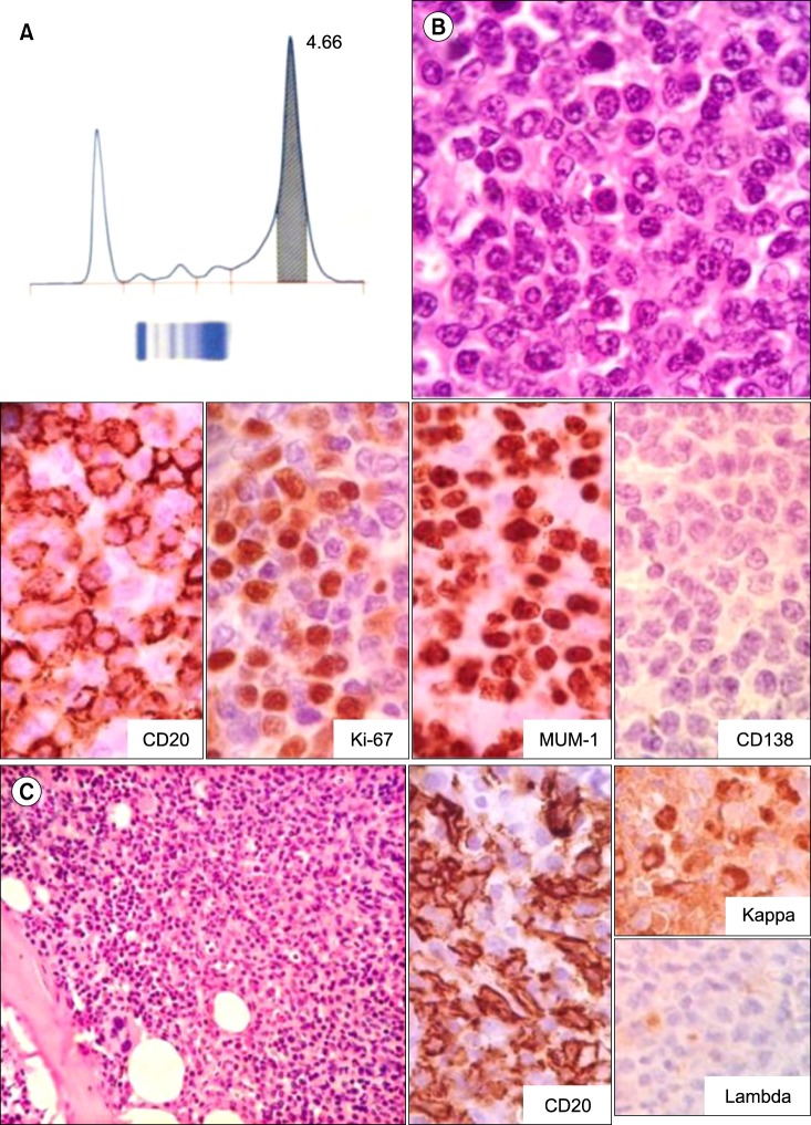
Fig. 1 (A) Serum protein electrophoresis showing an M band. (B) Lymph node biopsy showing large atypical lymphoid cells with irregular contours, brisk mitoses, and prominent nucleoli (hematoxylin and eosin, ×400), which were positive for CD20, MUM-1, and Ki-67 (70%) and negative for CD138. (C) Bone marrow biopsy showed lymphoid cells in a diffuse and nodular pattern (hematoxylin and eosin, ×200), which were CD20 and kappa positive and lambda negative.
Reference
-
1. Economopoulos T, Papageorgiou S, Pappa V, et al. Monoclonal gammopathies in B-cell non-Hodgkin's lymphomas. Leuk Res. 2003; 27:505–508. PMID: 12648510.
Article2. Chen M, Abedi M. Atypical lymphocytosis, cold agglutinin hemolytic anemia, and monoclonal gammopathy in an HIV patient with marrow involvement by diffuse large B-cell lymphoma. Blood. 2013; 122:3711. PMID: 24427808.
Article3. Charafeddine KM, Jabbour MN, Kadi RH, Daher RT. Extended use of serum free light chain as a biomarker in lymphoproliferative disorders: a comprehensive review. Am J Clin Pathol. 2012; 137:890–897. PMID: 22586047.
- Full Text Links
-
- Actions
-
Cited
- CITED
-
- Close
- Share
-
- Similar articles
-
- The Expression of p16 in Diffuse Large B-cell Lymphoma and Its Prognostic Implications
- Relapse of Ocular Lymphoma following Primary Testicular Diffuse Large B-cell Lymphoma
- A Case of Epstein-Barr Virus-Positive Diffuse Large B-Cell Lymphoma Occurring in Thyroid Gland
- Classification of Gastrointestinal B-cell Lymphoma and Expression of Cyclin D1, bcl-2, bcl-6, p53 Protein and PCNA
- Inverse Psoriasis Developed in a Patient with Diffuse Large B Cell Lymphoma
