Blood Res.
2015 Mar;50(1):54-55. 10.5045/br.2015.50.1.54.
Regenerating blasts masquerading as relapse in a patient with ALL following G-CSF therapy
- Affiliations
-
- 1Department of Pathology and Laboratory Medicine, Medanta-The Medicity, Gurgaon, India. sachdev05@gmail.com
- KMID: 1787914
- DOI: http://doi.org/10.5045/br.2015.50.1.54
Abstract
- No abstract available.
MeSH Terms
Figure
-
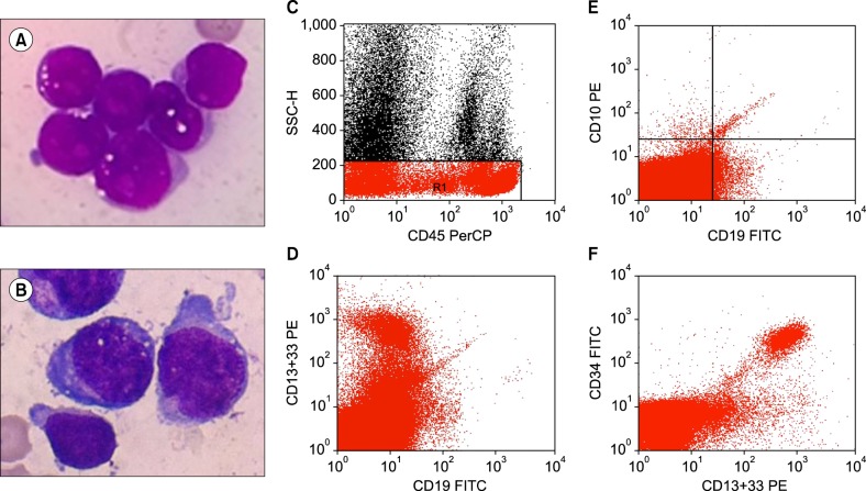
Fig. 1 (A) Diagnostic blasts with a high nucleocytoplasmic ratio and scant agranular cytoplasm (Giemsa stain, ×1,000). (B) Regenerating blasts are larger with moderate granular cytoplasm (Giemsa stain, ×1,000). (C-F) Immunophenotyping showing the regenerating blasts positive for CD45, CD34, CD13, and CD33 and negative for CD19 and CD10.
Reference
-
1. Liu CZ, Persad R, Inghirami G, et al. Transient atypical monocytosis mimic acute myelomonocytic leukemia in post-chemotherapy patients receiving G-CSF: report of two cases. Clin Lab Haematol. 2004; 26:359–362. PMID: 15485468.
Article2. Arici M, Hazendaroğlu IC, Erman M, Ozcebe O. Leukoerythroblastosis following the use of G-CSF. Am J Hematol. 1996; 52:123–124. PMID: 8638638.
Article
- Full Text Links
-
- Actions
-
Cited
- CITED
-
- Close
- Share
-
- Similar articles
-
- Proportions of Cells Expressing CD38-/CD34+, CD38+/CD34+, CD19+/CD34+, or CD13,33+/CD34+ in the Regenerating Bone Marrows During Complete Remission of Acute Leukemia or After Bone Marrow Transplantation
- Complete Remission Induced by Filgrastim (rhG-CSF) in the Case of Relapsed Acute Lymphoblastic Leukemia after Unrelated Cord Blood Transplantation
- Expression of Granulocyte Colony-Stimulating Factor Receptor (G-CSFR) and Clinical Correlates in Acute Leukemia
- Successful Treatment of Isolated Central Nervous System Relapse with Intrathecal Chemotherapy in an Adolescent with Acute Promyelocytic Leukemia
- Differential responses of CD34-positive acute myelogenous leukemic blasts to the costimulating effects of stem cell factor with GM-CSF and/or IL-3
