New classification of lingual arch form in normal occlusion using three dimensional virtual models
- Affiliations
-
- 1Graduate School of Clinical Dental Science, The Catholic University of Korea, Seoul, Korea.
- 2College of Medicine, The Catholic University of Korea, Seoul, Korea.
- 3Department of Postgraduate Studies, Universidad Autonoma del Paraguay, Asuncion, Paraguay.
- 4Postgraduate Orthodontic Program, Arizona School of Dentistry & Oral Health, A.T. Still University, Mesa, AZ, USA.
- 5Graduate School of Dentistry, Kyung Hee University, Seoul, Korea.
- 6Private Practice, Incheon, Korea.
- 7Department of Orthodontics, School of Dentistry, Dental Research Institute, Seoul National University, Seoul, Korea.
- 8Department of Orthodontics, Seoul St. Mary's Hospital, The Catholic University of Korea, Seoul, Korea. kook190036@yahoo.com
- KMID: 1787608
- DOI: http://doi.org/10.4041/kjod.2015.45.2.74
Abstract
OBJECTIVE
The purposes of this study were 1) to classify lingual dental arch form types based on the lingual bracket points and 2) to provide a new lingual arch form template based on this classification for clinical application through the analysis of three-dimensional virtual models of normal occlusion sample.
METHODS
Maxillary and mandibular casts of 115 young adults with normal occlusion were scanned in their occluded positions and lingual bracket points were digitized on the virtual models by using Rapidform 2006 software. Sixty-eight cases (dataset 1) were used in K-means cluster analysis to classify arch forms with intercanine, interpremolar and intermolar widths and width/depth ratios as determinants. The best-fit curves of the mean arch forms were generated. The remaining cases (dataset 2) were mapped into the obtained clusters and a multivariate test was performed to assess the differences between the clusters.
RESULTS
Four-cluster classification demonstrated maximum intercluster distance. Wide, narrow, tapering, and ovoid types were described according to the intercanine and intermolar widths and their best-fit curves were depicted. No significant differences in arch depths existed among the clusters. Strong to moderate correlations were found between maxillary and mandibular arch widths.
CONCLUSIONS
Lingual arch forms have been classified into 4 types based on their anterior and posterior dimensions. A template of the 4 arch forms has been depicted. Three-dimensional analysis of the lingual bracket points provides more accurate identification of arch form and, consequently, archwire selection.
Keyword
Figure
-
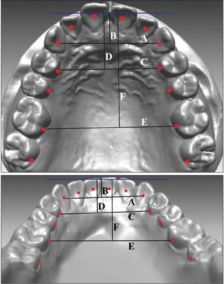
Figure 1 Lingual bracket points (red dots) on the three-dimensional virtual models. Arch dimensions were measured from these points. A, intercanine width; B, intercanine depth; C, interpremolar width; D, interpremolar depth; E, intermolar width; F, intermolar depth.
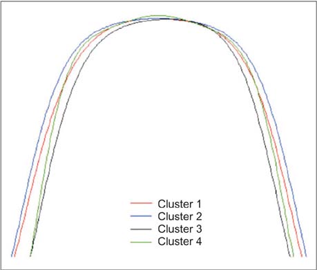
Figure 2 Superimposition of the best-fit curves of the mandibular arch clusters.
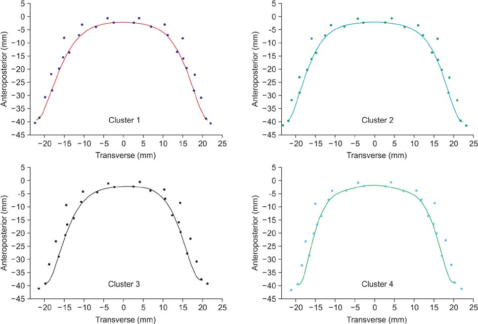
Figure 3 Template of mandibular lingual arch forms.
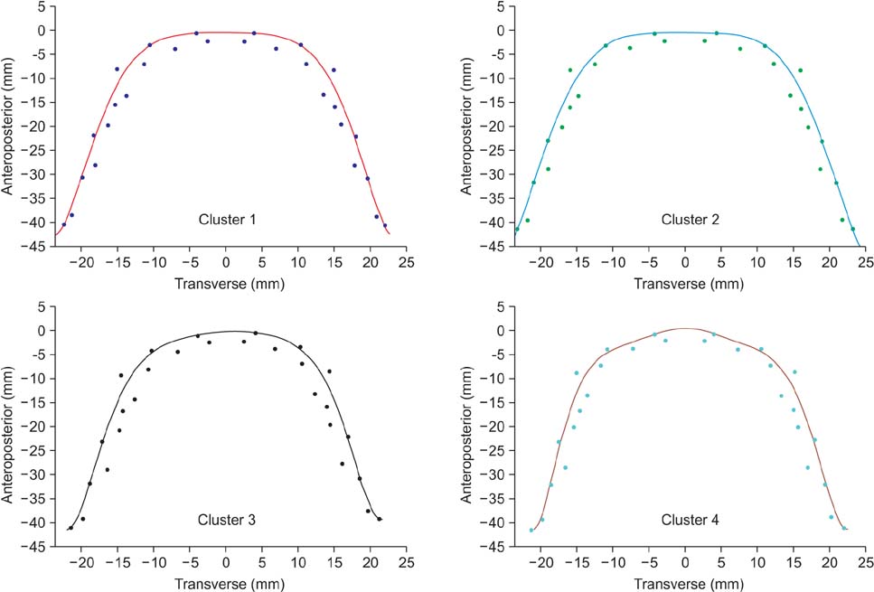
Figure 4 Template of maxillary lingual arch forms.
Cited by 5 articles
-
Frictional property comparisons of conventional and self-ligating lingual brackets according to tooth displacement during initial leveling and alignment: an in vitro mechanical study
Do-Yoon Kim, Bum-Soon Lim, Seung-Hak Baek
Korean J Orthod. 2016;46(2):87-95. doi: 10.4041/kjod.2016.46.2.87.Clustering of craniofacial patterns in Korean children with snoring
Stephanie Maritza Anderson, Hoi-Jeong Lim, Ki-Beom Kim, Sung-Wan Kim, Su-Jung Kim
Korean J Orthod. 2017;47(4):248-255. doi: 10.4041/kjod.2017.47.4.248.The effect of occlusogingival placement of clinical bracket points on the adaptation of a straight wire to the lingual arch form
Amir Hossein Abdi, Saeed Reza Motamedian, Ehsan Balaghi, Mahtab Nouri
Korean J Orthod. 2018;48(4):236-244. doi: 10.4041/kjod.2018.48.4.236.In-vitro investigation of the mechanical friction properties of a computer-aided design and computer-aided manufacturing lingual bracket system under diverse tooth displacement condition
Do-Yoon Kim, Sang-Woon Ha, Il-Sik Cho, Il-Hyung Yang, Seung-Hak Baek
Korean J Orthod. 2019;49(2):73-80. doi: 10.4041/kjod.2019.49.2.73.A novel classification of anterior alveolar arch forms and alveolar bone thickness: A cone-beam computed tomography study
Atcharee Bulyalert, Atiphan Pimkhaokham
Imaging Sci Dent. 2018;48(3):191-199. doi: 10.5624/isd.2018.48.3.191.
Reference
-
1. de la Cruz A, Sampson P, Little RM, Artun J, Shapiro PA. Long-term changes in arch form after orthodontic treatment and retention. Am J Orthod Dentofacial Orthop. 1995; 107:518–530.
Article2. Felton JM, Sinclair PM, Jones DL, Alexander RG. A computerized analysis of the shape and stability of mandibular arch form. Am J Orthod Dentofacial Orthop. 1987; 92:478–483.
Article3. Shapiro PA. Mandibular dental arch form and dimension. Treatment and postretention changes. Am J Orthod. 1974; 66:58–70.4. Nam HJ, Son WS, Park SB, Kim SS. Changes of mandibular dental arch during surgical-orthodontic treatment in skeletal class III malocclusion individuals. Korean J Orthod. 2008; 38:283–298.
Article5. Wheeler RC. A textbook of dental anatomy and physiology. Philadelphia: W. B. Saunders Company;1950. p. 196–215. p. 352–406.6. Lee KJ, Trang VT, Bayome M, Park JH, Kim Y, Kook YA. Comparison of mandibular arch forms of Korean and Vietnamese patients by using facial axis points on three-dimensional models. Korean J Orthod. 2013; 43:288–293.
Article7. Suk KE, Park JH, Bayome M, Nam YO, Sameshima GT, Kook YA. Comparison between dental and basal arch forms in normal occlusion and Class III malocclusions utilizing cone-beam computed tomography. Korean J Orthod. 2013; 43:15–22.
Article8. AlHarbi S, Alkofide EA, AlMadi A. Mathematical analyses of dental arch curvature in normal occlusion. Angle Orthod. 2008; 78:281–287.
Article9. Battagel JM. Individualized catenary curves: their relationship to arch form and perimeter. Br J Orthod. 1996; 23:21–28.
Article10. Bonwill WG, Huwley C. Individual arch form. St. Louis: CV Mosby;1966. p. 84–86.11. Braun S, Hnat WP, Fender DE, Legan HL. The form of the human dental arch. Angle Orthod. 1998; 68:29–36.12. Currier JH. A computerized geometric analysis of human dental arch form. Am J Orthod. 1969; 56:164–179.
Article13. Ferrario VF, Sforza C, Miani A Jr, Tartaglia G. Maxillary versus mandibular arch form differences in human permanent dentition assessed by Euclidean-distance matrix analysis. Arch Oral Biol. 1994; 39:135–139.
Article14. Ferrario VF, Sforza C, Miani A Jr, Tartaglia G. Mathematical definition of the shape of dental arches in human permanent healthy dentitions. Eur J Orthod. 1994; 16:287–294.
Article15. Pepe SH. Polynomial and catenary curve fits to human dental arches. J Dent Res. 1975; 54:1124–1132.
Article16. Sampson PD. Dental arch shape: a statistical analysis using conic sections. Am J Orthod. 1981; 79:535–548.
Article17. Raberin M, Laumon B, Martin JL, Brunner F. Dimensions and form of dental arches in subjects with normal occlusions. Am J Orthod Dentofacial Orthop. 1993; 104:67–72.
Article18. Chae JH, Song JW, Cha JY, Choi JS, Park YC. Labial and buccal surface contours of Korean normal occlusion in a three-dimensional digital model. Korean J Orthod. 2008; 38:95–103.
Article19. Leifert MF, Leifert MM, Efstratiadis SS, Cangialosi TJ. Comparison of space analysis evaluations with digital models and plaster dental casts. Am J Orthod Dentofacial Orthop. 2009; 136:16.e1–16.e4.
Article20. Zilberman O, Huggare JA, Parikakis KA. Evaluation of the validity of tooth size and arch width measurements using conventional and three-dimensional virtual orthodontic models. Angle Orthod. 2003; 73:301–306.21. Lim MY, Lim SH. Comparison of model analysis measurements among plaster model, laser scan digital model, and cone beam CT image. Korean J Orthod. 2009; 39:6–17.
Article22. Kim BI, Bayome M, Kim Y, Baek SH, Han SH, Kim SH, et al. Comparison of overjet among 3 arch types in normal occlusion. Am J Orthod Dentofacial Orthop. 2011; 139:e253–e260.
Article23. Kook YA, Bayome M, Park SB, Cha BK, Lee YW, Baek SH. Overjet at the anterior and posterior segments: three-dimensional analysis of arch coordination. Angle Orthod. 2009; 79:495–501.
Article24. Bayome M, Han SH, Choi JH, Kim SH, Baek SH, Kim DJ, et al. New clinical classification of dental arch form using facial axis points derived from three-dimensional models. Aust Orthod J. 2011; 27:117–124.25. Kim YL, Sung JH. A study on morphologic characteristics of lingual surface of crown and lingual archform of Korean adult with normal occlusion. Korean J Orthod. 1995; 25:209–221.26. Lombardo L, Saba L, Scuzzo G, Takemoto K, Oteo L, Palma JC, et al. A new concept of anatomic lingual arch form. Am J Orthod Dentofacial Orthop. 2010; 138:260.e1–260.e13.
Article27. Stamm T, Hohoff A, Ehmer U. A subjective comparison of two lingual bracket systems. Eur J Orthod. 2005; 27:420–426.
Article28. Caniklioglu C, Oztürk Y. Patient discomfort: a comparison between lingual and labial fixed appliances. Angle Orthod. 2005; 75:86–91.29. Triviño T, Siqueira DF, Scanavini MA. A new concept of mandibular dental arch forms with normal occlusion. Am J Orthod Dentofacial Orthop. 2008; 133:10.e15–10.e22.
Article30. Noroozi H, Nik TH, Saeeda R. The dental arch form revisited. Angle Orthod. 2001; 71:386–389.
- Full Text Links
-
- Actions
-
Cited
- CITED
-
- Close
- Share
-
- Similar articles
-
- A study on morphologic characteristics of lingual surface of crown and lingual archform of Korean adult with normal occlusion
- The dental arch form in normal occlusion
- A morphologic characteristics study on crown of lingual surface with normal occlusion in Korean adults
- A study on the dental arch by occlusogram in normal occlusion
- Three dimensional structural analysis between dental arch and basal bone in normal occlusion
