J Korean Med Sci.
2013 Apr;28(4):614-619. 10.3346/jkms.2013.28.4.614.
Clinical Characteristics of 75 Patients with Leukemia Cutis
- Affiliations
-
- 1Department of Dermatology, Seoul St. Mary's Hospital, Seoul, Korea. yymmpark6301@hotmail.com
- 2Department of Dermatology, Incheon St. Mary's Hospital, Incheon, Korea.
- 3Department of Dermatology, Yeouido St. Mary's Hospital, College of Medicine, The Catholic University of Korea, Seoul, Korea.
- KMID: 1786972
- DOI: http://doi.org/10.3346/jkms.2013.28.4.614
Abstract
- Leukemia cutis (LC) is defined as a neoplastic leukocytic infiltration of the skin. Few clinical studies are available on recent trends of LC in Korea. The purpose of this study was to analyze the clinical features and prognosis of LC in Korea and to compare findings with previous studies. We performed a retrospective study of 75 patients with LC and evaluated the patients' age and sex, clinical features and skin lesion distribution according to the type of leukemia, interval between the diagnosis of leukemia and the development of LC, and prognosis. The male to female ratio was 2:1, and the mean age at diagnosis was 37.6 yr. The most common cutaneous lesions were nodules. The most commonly affected site was the extremities in acute myelocytic leukemia and chronic myelocytic leukemia except for acute lymphocytic leukemia. Compared with previous studies, there was an increasing tendency in the proportion of males and nodular lesions, and LC most often occurred in the extremities. The prognosis of LC was still poor within 1 yr, which was similar to the results of previous studies. These results suggest that there is a difference in the clinical characteristics and predilection sites according to type of leukemia.
Keyword
MeSH Terms
Figure
-
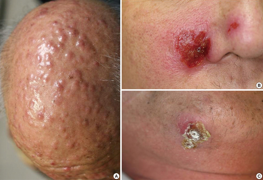
Fig. 1 Gross manifestation on face. (A) Asymptomatic, multiple, variable-sized, erythematous papules on the face. (B) Painful, oozing, erythematous, erosive patches on the cheek. (C) Tender, crusted plaque on the chin.
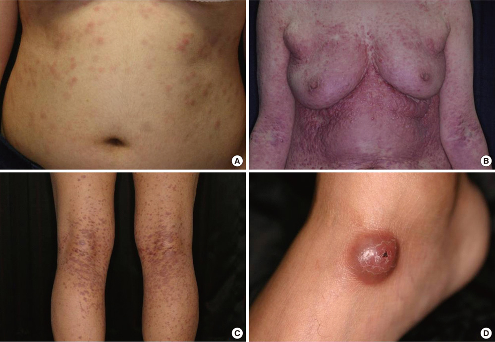
Fig. 2 Gross skin lesions on the trunk or extremities. (A) Multiple, erythematous nodules on the trunk. (B) Multiple, erythematous nodules on the whole body. (C) Pruritic, erythematous papules on both extremities. (D) Solitary, reddish-brown mass on the ankle.
Reference
-
1. Costello MJ, Canizares O, Montague M, Buncke CM. Cutaneous manifestations of myelogenous leukemia. AMA Arch Derm. 1955. 71:605–614.2. Wagner G, Fenchel K, Back W, Schulz A, Sachse MM. Leukemia cutis - epidemiology, clinical presentation, and differential diagnoses. J Dtsch Dermatol Ges. 2012. 10:27–36.3. Cho KH, Jeon HP, Kim JA, Lee SK, Park SH, Kim BK. A clinicopathological study of leukemia cutis. Korean J Dermatol. 1990. 28:321–330.4. Jang IG, Lee DW, Han CW, Kim CC, Cho BK. A clinical observation on leukemia cutis. Korean J Dermatol. 1996. 34:507–514.5. Jang KA, Chi DH, Choi JH, Sung KJ, Moon KC, Koh JK. Leukemia cutis: a clinico-pathologic study of 23 patients. Korean J Dermatol. 2000. 38:15–22.6. Kaddu S, Zenahlik P, Beham-Schmid C, Kerl H, Cerroni L. Specific cutaneous infiltrates in patients with myelogenous leukemia: a clinicopathologic study of 26 patients with assessment of diagnostic criteria. J Am Acad Dermatol. 1999. 40:966–978.7. Bakst RL, Tallman MS, Douer D, Yahalom J. How I treat extramedullary myeloid leukemia. Blood. 2011. 118:3785–3793.8. Braverman IM. Skin signs of systemic disease. 1981. 3rd ed. Philadelphia: Saunders;179–196.9. Su WP. Clinical, histopathologic, and immunohistochemical correlations in leukemia cutis. Semin Dermatol. 1994. 13:223–230.10. Choi HS, Hahn JS, Lee KH, Cho SH. A case of leukemia cutis associated with B-cell chronic lymphocytic leukemia. Korean J Hematol. 2003. 38:147–150.11. Moon TK, Lee BJ, Lee SH, Ahn SK, Lee WS. Leukemic macrocheilitis associated with chronic lymphocytic leukemia. Korean J Dermatol. 1994. 32:1114–1118.12. Weinstein HJ. Wyngaarden JB, Smith LH, editors. The acute leukemias. The text book of medicine. 1998. 18th ed. Philadelphia: W. B. Saunders;1001–1009.13. Watson KM, Mufti G, Salisbury JR, du Vivier AW, Creamer D. Spectrum of clinical presentation, treatment and prognosis in a series of eight patients with leukaemia cutis. Clin Exp Dermatol. 2006. 31:218–221.14. Ohno S, Yokoo T, Ohta M, Yamamoto M, Danno K, Hamato N, Tomii K, Ohno Y, Kobashi Y. Aleukemic leukemia cutis. J Am Acad Dermatol. 1990. 22:374–377.15. Su WP, Buechner SA, Li CY. Clinicopathologic correlations in leukemia cutis. J Am Acad Dermatol. 1984. 11:121–128.16. Shaikh BS, Frantz E, Lookingbill DP. Histologically proven leukemia cutis carries a poor prognosis in acute nonlymphocytic leukemia. Cutis. 1987. 39:57–60.17. Paydaş S, Zorludemir S. Leukaemia cutis and leukaemic vasculitis. Br J Dermatol. 2000. 143:773–779.18. Cho-Vega JH, Medeiros LJ, Prieto VG, Vega F. Leukemia cutis. Am J Clin Pathol. 2008. 129:130–142.
- Full Text Links
-
- Actions
-
Cited
- CITED
-
- Close
- Share
-
- Similar articles
-
- A Case of Aleukemic Leukemia Cutis Occurring During Spontaneous Remission of Acute Myelomonocytic Leukemia
- A Case of Leukemia Cutis Associated with B-cell Chronic Lymphocytic Leukemia
- A Case of Leukemia Cutis Showing Rosacea-like Cutaneous Lesions
- A Clinicopathological Study of Leukemia Cutis
- Two Cases of Leukemia Cutis Showing Clinical Improvement with Systemic Retinoid Administration
