J Korean Med Sci.
2013 Mar;28(3):480-484. 10.3346/jkms.2013.28.3.480.
Diffuse Follicular Variant of Papillary Thyroid Carcinoma in a 69-Year-old Man with Extensive Extrathyroidal Extension: A Case Report
- Affiliations
-
- 1Department of Pathology, Gangnam Severance Hospital, Yonsei University College of Medicine, Seoul, Korea. soonwonh@yuhs.ac
- 2Department of Surgery, Gangnam Severance Hospital, Yonsei University College of Medicine, Seoul, Korea.
- KMID: 1786950
- DOI: http://doi.org/10.3346/jkms.2013.28.3.480
Abstract
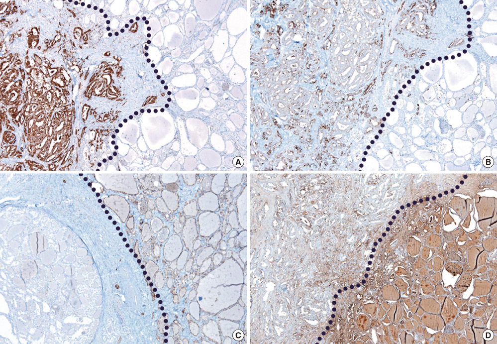
- Diffuse follicular variant papillary thyroid carcinoma (DFVPTC) is a rare variant papillary thyroid carcinoma. DFVPTC typically occurs in young females, extensively involves one lobe or both lobes entirely with frequent nodal metastasis and vascular invasion. In contrast to the other subtypes of follicular variant, DFVPTC has biologically aggressive behavior. We present a case of DFVPTC arising in a 69-yr-old male patient. He presented hoarseness for a few months. Following diagnosis of malignancy on aspiration cytology, total thyroidectomy with neck dissection was performed. The tumor involved both lobes of thyroid, encroaching the surrounding structures including tracheal cartilage and esophagus. Multiple lymph node metastasis and vascular invasion were also found. The patient passed away due to the unexplained bleeding of surgical site.
Keyword
MeSH Terms
-
Aged
Antigens, CD56/metabolism
Carcinoma, Papillary, Follicular/*diagnosis/pathology/surgery
Galectin 3/metabolism
Humans
Immunohistochemistry
Keratin-19/metabolism
Lymph Nodes/pathology
Lymphatic Metastasis
Male
Thyroid Neoplasms/*diagnosis/pathology/surgery
Thyroidectomy
Tomography, X-Ray Computed
Antigens, CD56
Galectin 3
Keratin-19
Figure
Reference
-
1. Schlumberger MJ. Papillary and follicular thyroid carcinoma. N Engl J Med. 1998. 338:297–306.2. Nikiforov Y. Diagnostic pathology and molecular genetics of the thyroid. 2009. Baltimore: Lippincott William & Wilkins;176–183.3. Sobrinho-Simões M, Soares J, Carneiro F, Limbert E. Diffuse follicular variant of papillary carcinoma of the thyroid: report of eight cases of a distinct aggressive type of thyroid tumor. Surg Pathol. 1990. 3:189–203.4. Mizukami Y, Nonomura A, Michigishi T, Ohmura K, Noguchi M, Ishizaki T. Diffuse follicular variant of papillary carcinoma of the thyroid. Histopathology. 1995. 27:575–577.5. Ivanova R, Soares P, Castro P, Sobrinho-Simões M. Diffuse (or multinodular) follicular variant of papillary thyroid carcinoma: a clinicopathologic and immunohistochemical analysis of ten cases of an aggressive form of differentiated thyroid carcinoma. Virchows Arch. 2002. 440:418–424.
- Full Text Links
- Actions
-
Cited
- CITED
-
- Close
- Share
- Similar articles
-
- Follicular Variant of Papillary Carcinoma Thyroid with Massive Angioinvasion of the Internal Jugular Vein: Our Approach
- Diffuse Sclerosing Variant of Papillary Thyroid Carcinoma: Case Report
- A Case of Mixed Follicular-Papillary Thyroid Carcinoma
- Sonographic Index for Extrathyroidal Extension of Papillary Thyroid Carcinoma
- Differential Diagnosis of a Follicular Carcinoma and Papillary Carcinoma of the Thyroid Gland Based on Sonographic Findings


