J Korean Med Sci.
2004 Feb;19(1):101-106. 10.3346/jkms.2004.19.1.101.
The Relationship between Disc Degeneration and Morphologic Changes in the Intervertebral Foramen of the Cervical Spine: A Cadaveric MRI and CT Study
- Affiliations
-
- 1Department of Orthopaedic Surgery, Chosun University Hospital, Gwangju, Korea. jwyou@chosun.ac.kr
- KMID: 1785702
- DOI: http://doi.org/10.3346/jkms.2004.19.1.101
Abstract
- A cadaveric study was performed to investigate the relationship between disc degeneration and morphological changes in the intervertebral foramen of cervical spine, including the effect on the nerve root. Seven fresh frozen human cadavers were dissected from C1 to T1, preserving the ligaments, capsules, intervertebral disc and the neural structures. The specimens were scanned with MRI and then scanned through CT scan in the upright position. Direct mid-sagittal and 45 degree oblique images were obtained to measure the dimension of the intervertebral disc height, foraminal height, width, area and segmental angles. Disc degeneration was inversely correlated with disc height. There was a significant correlation between disc degeneration and foraminal width (p<0.005) and foraminal area (p< 0.05), but not with foraminal height. Disc height was correlated with foraminal width but not with height. The segmental angles were decreased more in advanced degenerated discs. There was a correlation between nerve root compression and decreased foraminal width and area (p<0.005). This information and critical dimensions of the intervertebral foramen for nerve root compression should help in the diagnosis of foraminal stenosis of the cervical spine in patients presenting with cervical spondylosis and radiculopathy.
Keyword
MeSH Terms
Figure
-
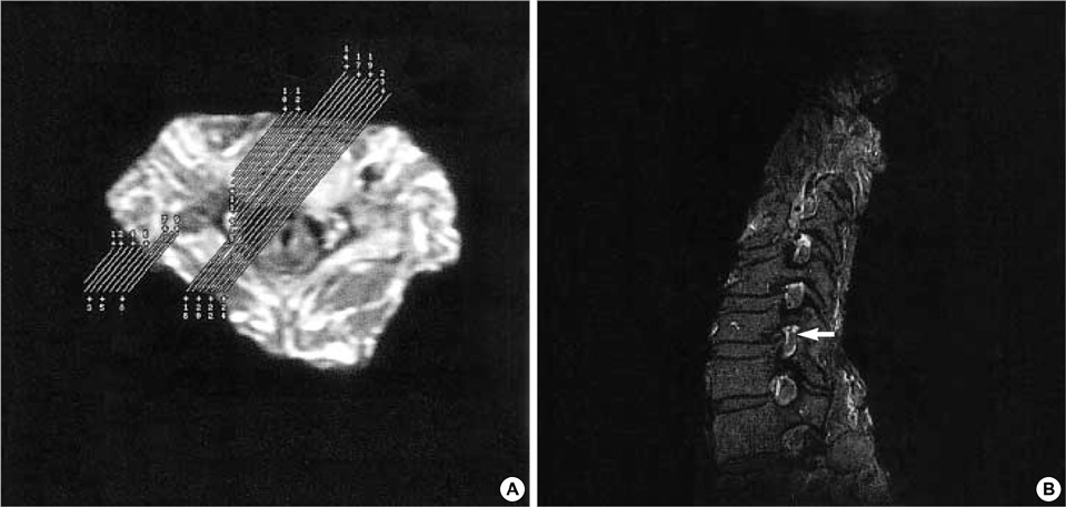
Fig. 1 (A) T1-weighted (500/16), 45 degree oblique projections of magnetic resonance imaging of the cervical spine, 1.0 mm slice thickness. (B) MRI shows decreased width and area at C5-6 intervertebral foramen (arrow), but relative conservation of foraminal height.
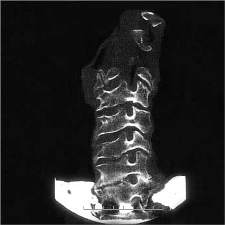
Fig. 2 Parasagittal oblique plane CT images of the cervical spine.
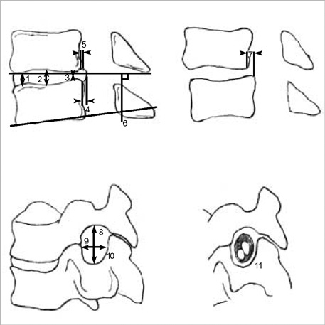
Fig. 3 Diagram showing the sagittal parameters of measurement. 1. Anterior disc height, 2. Midpoint disc height, 3. Posterior disc height, 4. Posterior bulging of disc, 5. Distance of spur, 6. Segmental angle of body, 7. Vertebral body translation, 8. Foraminal height, 9. Middle foraminal width, 10. Foraminal area, 11. Nerve root condition.
Reference
-
1. Resnick D. Degenerative diseases of the vertebral column. Radiology. 1985. 156:3–14.
Article2. Kirkaldy-Willis WH, Wedge JH, Yong-Hing K, Reilly J. Pathology and pathogenesis of lumbar spondylosis and stenosis. Spine. 1978. 3:319–328.
Article3. An HS, Glover JM. Lumbar spinal stenosis. Historical perspective, classification and pathoanatomy. Sem Spin Surg. 1994. 6:67–77.4. Rauschning W. Normal and pathologic anatomy of the lumbar root canals. Spine. 1987. 12:1008–1019.
Article5. Kunogi J, Hasue M. Diagnosis and operative treatment of intraforaminal and extraforaminal nerve root compression. Spine. 1991. 16:1312–1320.
Article6. Sato K, Kikuchi S. An anatomic study of foraminal nerve root lesions in the lumbar. Spine. 1993. 18:2246–2251.7. Lejeune JP, Hladky JP, Cotten A, Vinchon M, Christiaens JL. Foraminal lumbar disc herniation. Experience with 83 patients Spine. 1994. 19:1905–1908.
Article8. Frymoyer JW, Gordon SL. New perspectives on low back pain. 1989. Park Ridge, Illinois: American Academy of Orthopaedic Surgeons;191–193.9. Gilad I, Nissan M. A study of vertebra and disc geometric relations of the human cervical and lumbar spine. Spine. 1986. 11:154–157.
Article10. Kameyama T, Hashizume Y, Ando T, Takahashi A. Morphometry of the normal cadaveric cervical spinal cord. Spine. 1994. 19:2077–2081.
Article11. Okada Y, Ikata T, Katoh S, Yamada H. Morphologic analysis of the cervical spinal cord, dural tube, and spinal canal by magnetic resornance imaging in normal adults and patients with cervical spondylotic myelopathy. Spine. 1994. 19:2331–2335.12. Panjabi MM, Duranceau J, Goel V, Oxland T, Takata K. Cervical human vertebrae. Quantitative three-dimensional anatomy of the middle and lower regions. Spine. 1991. 16:861–869.
Article13. Pech P. Corrective investigation of craniospinal anatomy and pathology with computed tomography, magnetic resonance imaging and cryomicrotomy. Acta Radiol. 1988. 372:Suppl. 127–148.14. Yenerich DO, Haughton VM. Oblique plane MR imaging of the cervical spine. J Comput Assist Tomogr. 1986. 10:823–826.
Article15. Schnebel B, Kingston S, Watkins R, Dillin W. Comparison of MRI to contrast CT in the diagnosis of spinal stenosis. Spine. 1989. 14:332–337.
Article16. Modic MT, Masaryk TJ, Ross JS, Mulopulos GP, Bundschuh CV, Bohlman H. Cervical radioculopathy: value of oblique MR imaging. Radiology. 1987. 163:227–231.17. Tsurda JS, Norman D, Dillon W, Newton TH, Mills DG. Three-dimensional gradient-recalled MR imaging as a screening tool for the diagnosis of cervical radiculopathy. AJNR Am J Neuroradiol. 1989. 10:1263–1271.18. Yousem DM, Atlas SW, Goldberg HI, Grossman RI. Degenerative narrowing of the cervical spine neural foramina: evaluation with high resolution 3DFT gradient-echo MR imaging. AJNR Am J Neuroradiol. 1991. 12:229–236.19. Ross JS, Ruggieri PM, Glicklich M, Obuchowski N, Dillinger J, Masaryk TJ, Qu Y, Modic MT. 3D MRI of the cervical spine: Low flip angle FISP vs. Gd-DTPA TurboFLASH in degenerative disk disease. J Comput Assist Tomogr. 1997. 17:26–33.20. Ebraheim NA, An HS, Xu R, Ahmad M, Yeasting RA. The quantitative anatomy of the cervical nerve root groove and the intervertebral foramen. Spine. 1996. 21:1619–1623.
Article21. Macnab I. The traction spur. J Bone Joint Surg. 1971. 53-A:663–670.
Article22. Panjabi MM, Takata K, Goel VK. Kinematics of lumbar intervertebral foramen. Spine. 1983. 8:348–357.
Article23. Penning L, Wilmink JT. Posture-dependent bilateral compression of L4 or L5 nerve roots in facet hypertrophy: A dynamic CT-myelographic study. Spine. 1987. 12:488–500.24. Schönström N, Lindahl S, Willén J, Hansson T. Dynamic changes in the dimensions of the lumbar spinal canal: An experimental study in vitro. J Orthop Res. 1989. 7:115–121.
Article25. Yoo JU, Zou D, Edwards WT, Bayley J, Yuan HA. Effect of cervical spine motion on the neuroforaminal dimensions of human cervical spine. Spine. 1992. 17:1131–1136.
Article26. Farmer JC, Wisneski RJ. Cervical spine nerve root compression. An analysis of neuroforaminal pressures with varying head and arm positions. Spine. 1994. 19:1850–1855.
- Full Text Links
-
- Actions
-
Cited
- CITED
-
- Close
- Share
-
- Similar articles
-
- Biochemical Factors of Intervertebral Disc Degeneration: Implications for Disc Regeneration
- Effect of Facet Tropism on the Degeneration of the Cervical Facet Joint and Intervertebral Disc
- Correlation of Cervical Disc Degeneration with Sagittal Alignments of Cervical Spine
- The Actual Level of Symptomatic Soft Disc Herniation in Patients with Cervical Disc Herniation
- Clinical Relationship of Degenerative Changes between the Cervical and Lumbar Spine
