Plain Radiologic Findings and Chronological Changes of Incipient Phase Osteosarcoma Overlooked by Primary Physicians
- Affiliations
-
- 1Department of Orthopedic Surgery, Korea Cancer Center Hospital, Seoul, Korea. dgjeon@kcch.re.kr
- KMID: 1784672
- DOI: http://doi.org/10.4055/cios.2014.6.2.230
Abstract
- BACKGROUND
We assessed the plain radiographic characteristics of 10 cases of osteosarcomas during the initial painful period that had been overlooked by a primary physician. In addition, we evaluated chronologic changes in radiographic findings from initial symptomatic period to the time of accurate diagnosis.
METHODS
The clinical records were reviewed for clinical parameters including age, sex, location, presenting symptoms, initial diagnosis, duration from initial symptoms to definite diagnosis, and initial and follow-up plain radiographic findings of the lesion.
RESULTS
Initial clinical diagnoses included a sprain in 6, growing pain in 2, stress fracture in 1, and infection in 1 patient. Initial plain radiographic findings were trabecular destruction (100%), cortical disruption (60%), periosteal reaction (60%), and soft tissue mass (10%). Intramedullary matrix changes were osteosclerosis in 6 and osteolysis in 4 patients. On progression, 4 cases with minimal sclerosis changed to osteoblastic lesion in 3 patients and osteolytic lesion in 1. Four cases with faint osteolytic foci transformed into osteolytic lesion in 3 and mixed pattern in 1.
CONCLUSIONS
Notable plain radiologic findings of incipient-stage osteosarcoma include trabecular disruption along with faint osteosclerosis or osteolysis. In symptomatic patients with trabecular destruction, additional imaging study including magnetic resonance imaging should be performed to exclude osteosarcoma in the incipient phase, even without radiologic findings suggesting malignant tumor, such as cortical destruction or periosteal reaction.
MeSH Terms
Figure
-
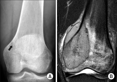
Fig. 1 An 18-year-old boy presented with pain at distal femur for the prior 3 weeks (case 10). (A) The plain radiograph demonstrated a faint sclerotic change in the metaphysis of the distal femur (arrow). (B) A T1-weighted fat suppression gadolinium-enhanced magnetic resonance imaging scan taken 1 day later demonstrated a heterogeneously enhancing lesion with an extraosseous tumor mass.
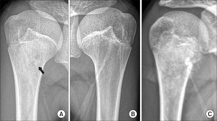
Fig. 2 A 14-year-old girl presented with pain at the right shoulder (case 6). (A) The plain radiograph showed trabecular disruption with minimal sclerotic change in the metaphysis of the proximal humerus (arrow). (B) A plain radiograph of the left humerus, taken simultaneously, showed intact trabeculae. (C) A plain radiograph taken 8 weeks later clearly showed a destructive osteoblastic lesion with an extraosseous lesion.

Fig. 3 A 7-year-old girl presented with night pain at the distal femur (case 1). (A) The plain radiograph showed minimal cortical disruption with a moderate sclerotic change in the metaphysis of the distal femur (arrow). (B) A week later, the patient returned with a fracture after a minor traumatic event. (C) A plain radiograph taken 4 months after closed reduction and pinning showed a destructive lesion with a huge soft tissue mass.

Fig. 4 A 12-year-old girl presented with pain after exercise (case 5). (A) The plain radiograph showed focal cortical breakage and faint sclerosis in the proximal tibia (arrow). (B) Under the assumption of a stress fracture, the primary physician recommended immobilization for 4 weeks; the plain radiograph showed increased sclerosis and a periosteal reaction. (C) At 14 weeks, the patient still complained of pain and developed swelling. The plain radiograph clearly showed an extraosseous mass. (D) A T1-weighted fat suppression gadolinium-enhanced magnetic resonance imaging scan taken at this time demonstrated that the lesion was not a stress fracture but instead was an osteosarcoma.
Reference
-
1. Sneppen O, Hansen LM. Presenting symptoms and treatment delay in osteosarcoma and Ewing's sarcoma. Acta Radiol Oncol. 1984; 23(2-3):159–162.2. Widhe B, Widhe T. Initial symptoms and clinical features in osteosarcoma and Ewing sarcoma. J Bone Joint Surg Am. 2000; 82(5):667–674.3. Kim MS, Cho WH, Song WS, Lee SY, Jeon DG. time dependency of prognostic factors in patients with stage II osteosarcomas. Clin Orthop Relat Res. 2007; 463:157–165.4. Jeon DG, Lee SY, Cho WH, Song WS, Park JH. Primary osteosarcoma in patients older than 40 years of age. J Korean Med Sci. 2006; 21(4):715–718.5. de Santos LA, Edeiken BS. Subtle early osteosarcoma. Skeletal Radiol. 1985; 13(1):44–48.6. Andresen KJ, Sundaram M, Unni KK, Sim FH. Imaging features of low-grade central osteosarcoma of the long bones and pelvis. Skeletal Radiol. 2004; 33(7):373–379.7. Bertoni F, Unni KK, McLeod RA, Dahlin DC. Osteosarcoma resembling osteoblastoma. Cancer. 1985; 55(2):416–426.8. Tsuneyoshi M, Dorfman HD. Epiphyseal osteosarcoma: distinguishing features from clear cell chondrosarcoma, chondroblastoma, and epiphyseal enchondroma. Hum Pathol. 1987; 18(6):644–651.9. Rosenberg ZS, Lev S, Schmahmann S, Steiner GC, Beltran J, Present D. Osteosarcoma: subtle, rare, and misleading plain film features. AJR Am J Roentgenol. 1995; 165(5):1209–1214.10. Kim MS, Lee SY, Cho WH, et al. Prognostic effects of doctor-associated diagnostic delays in osteosarcoma. Arch Orthop Trauma Surg. 2009; 129(10):1421–1425.11. Jeon DG, Lee SY, Kim JW. Bone primary sarcomas undergone unplanned intralesional procedures - the possibility of limb salvage and their oncologic results. J Surg Oncol. 2006; 94(7):592–598.12. Kim MS, Lee SY, Cho WH, et al. Relationships between plain-film radiographic patterns and clinicopathologic variables in AJCC stage II osteosarcoma. Skeletal Radiol. 2008; 37(11):997–1001.13. Ayerza MA, Muscolo DL, Aponte-Tinao LA, Farfalli G. Effect of erroneous surgical procedures on recurrence and survival rates for patients with osteosarcoma. Clin Orthop Relat Res. 2006; 452:231–235.
