J Vet Sci.
2014 Jun;15(2):187-193. 10.4142/jvs.2014.15.2.187.
Computed tomographic evaluation of cervical vertebral canal and spinal cord morphometry in normal dogs
- Affiliations
-
- 1College of Veterinary Medicine and the Research Institute for Veterinary Science, Seoul National University, Seoul 151-742, Korea. heeyoon@snu.ac.kr
- 2College of Veterinary Medicine, Chonnam National University, Gwangju 500-757, Korea.
- KMID: 1784638
- DOI: http://doi.org/10.4142/jvs.2014.15.2.187
Abstract
- The height, width, and cross-sectional area of the vertebral canal and spinal cord along with the area ratio of spinal cord to vertebral canal in the cervical vertebra were evaluated in images obtained using computed tomography (CT). Measurements were taken at the cranial, middle, and caudal point of each cervical vertebra in eight clinically normal small breed dogs (two shih tzu, two miniature schnauzers, and four mixed breed), 10 beagles, and four German shepherds. CT myelography facilitated the delineation of the epidural space, subarachnoid space, and spinal cord except at the caudal portion of the 7th cervical vertebra. The spinal cord had a tendency to have a clear ventral border in the middle portion of the vertebral canal and lateral borders near both end plates. The height, width, and area of the vertebral canal and spinal cord in the cervical vertebra were increased as the size of dog increased. However, the ratio of the spinal cord area to vertebral canal area in the small dogs was higher than that of the larger dogs. Results of the present study could provide basic and quantitative information for CT evaluation of pathologic lesions in the cervical vertebra and spinal cord.
MeSH Terms
Figure
-
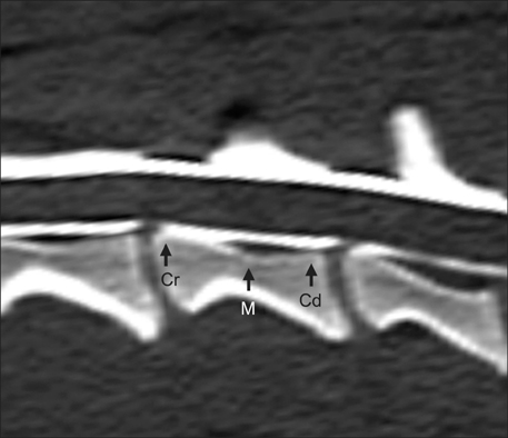
Fig. 1 Reformatted sagittal image of the fourth cervical vertebra derived from the initial transverse image. Arrows indicate the cranial (Cr), middle (M), and caudal (Cd) points that were used to measure the height, width, and area of the spinal cord and vertebral canal.
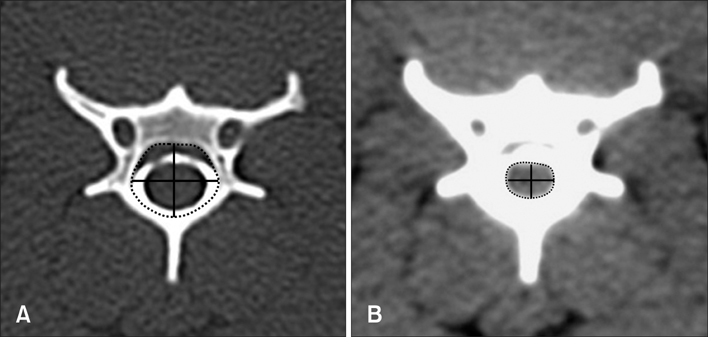
Fig. 2 Height, width, and area of the vertebral canal and spinal cord in the middle portion of the fourth cervical vertebra were measured on the images in which the vertebra window (A) and vertebra window (B) were manipulated.
Cited by 1 articles
-
Novel vertebral computed tomography indices in normal and spinal disorder dogs
Jongsu Lim, Youngmin Yoon, Taesung Hwang, Hee Chun Lee
J Vet Sci. 2018;19(2):296-300. doi: 10.4142/jvs.2018.19.2.296.
Reference
-
1. da Costa RC, Parent JM, Partlow G, Dobson H, Holmberg DL, LaMarre J. Morphologic and morphometric magnetic resonance imaging features of Doberman Pinschers with and without clinical signs of cervical spondylomyelopathy. Am J Vet Res. 2006; 67:1601–1612.
Article2. de Decker S, Gielen IMVL, Duchateau L, Polis I, van Bree HJJ, van Ham LML. Agreement and repeatability of linear vertebral body and canal measurements using computed tomography (CT) and low field magnetic resonance imaging (MRI). Vet Surg. 2010; 39:28–34.
Article3. de Decker S, Gielen IMVL, Duchateau L, Saunders JHH, van Bree HJJ, Polis I, van Ham LML. Magnetic resonance imaging vertebral canal and body ratios in Doberman Pinschers with and without disk-associated cervical spondylomyelopathy and clinically normal English Foxhounds. Am J Vet Res. 2011; 72:1496–1504.
Article4. Drost WT, Love NE, Berry CR. Comparison of radiography, myelography and computed tomography for the evaluation of canine vertebral and spinal cord tumors in sixteen dogs. Vet Radiol Ultrasound. 1996; 37:28–33.
Article5. Feeney DA, Evers P, Fletcher TF, Hardy RM, Wallace LJ. Computed tomography of the normal canine lumbosarcral spine: a morphologic perspective. Vet Radiol Ultrasound. 1996; 37:399–411.
Article6. Fourie SL, Kirberger RM. Relationship of cervical spinal cord diameter to vertebral dimensions: a radiographic study of normal dogs. Vet Radiol Ultrasound. 1999; 40:137–143.
Article7. Harris JH Jr. Radiographic evaluation of spinal trauma. Orthop Clin North Am. 1986; 17:75–86.
Article8. Haughton VM, Syvertsen A, Williams AL. Soft-tissue anatomy within the spinal canal as seen on computed tomography. Radiology. 1980; 134:649–655.
Article9. Hecht S, Huerta MM, Reed RB. Magnetic resonance imaging (MRI) spinal cord and canal measurements in normal dogs. Anat Histol Embryol. 2014; 43:36–41.
Article10. Itoh T, Nishimura R, Matsunaga S, Kadosawa T, Mochizuki M, Sasaki N. Syringomyelia and hydrocephalus in a dog. J Am Vet Med Assoc. 1996; 209:934–936.11. Ketonen L, Gyldensted C. Lumbar disc disease evaluated by myelography and postmyelography spinal computed tomography. Neuroradiology. 1986; 28:144–149.
Article12. Lamb CR. Common difficulties with myelographic diagnosis of acute intervertebral disc prolapse in the dog. J Small Anim Pract. 1994; 35:549–558.
Article13. Lee BC, Kazam E, Newman AD. Computed tomography of the spine and spinal cord. Radiology. 1978; 128:95–102.
Article14. Lee J, Williams B. Cervical vertebrae measurements in syringomyelia. Clin Radiol. 1977; 28:395–400.
Article15. Lim C, Kweon OK, Choi MC, Choi J, Yoon J. Computed tomographic characteristics of acute thoracolumbar intervertebral disc disease in dogs. J Vet Sci. 2010; 11:73–79.
Article16. Morgan JP, Ackerman N, Bailey CS, Pool RR. Vertebral tumors in the dog: a clinical radiologic, and pathologic study of 61 primary and secondary lesions. Veterinary Radiology. 1980; 21:197–212.
Article17. Morgan JP, Atilola M, Bailey CS. Vertebral canal and spinal cord mensuration: a comparative study of its effect on lumbosacral myelography in the Dachshund and German shepherd dog. J Am Vet Med Assoc. 1987; 191:951–957.18. Morgan JP, Miyabayashi T, Choy S. Cervical spine motion: radiographic study. Am J Vet Res. 1986; 47:2165–2169.19. Pavlov H, Torg JS, Robie B, Jahre C. Cervical spinal stenosis: determination with vertebral body ratio method. Radiology. 1987; 164:771–775.
Article20. Penning L, Wilmink JT, van Woerden HH, Knol E. CT myelographic findings in degenerative disorders of the cervical spine: clinical significance. AJR Am J Roentgenol. 1986; 146:793–801.
Article21. Sande RD. Radiography, myelography, computed tomography and magnetic imaging of the spine. Vet Clin North Am Small Anim Pract. 1992; 22:811–831.
Article22. Stanley JH, Schabel SI, Frey GD, Hungerford GD. Quantitative analysis of the cervical spinal canal by computed tomography. Neuroradiology. 1986; 28:139–143.
Article23. Tadmor R, Cacayorin ED, Kieffer SA. Advantages supplementary CT in myelography of intraspinal masses. AJNR Am J Neuroradiol. 1983; 4:618–621.24. Tidwell AS, Solano M, Schelling SH. Pediatric neuroimaging. Semin Vet Med Surg (Small Anim). 1994; 9:68–85.25. Wortman JA. Principles of X-ray computed tomography and magnetic resonance imaging. Semin Vet Med Surg (Small Anim). 1986; 1:176–184.26. Yu YL. Management of cervical spondylitic myelopathy. Lancet. 1984; 2:170–171.
Article
- Full Text Links
-
- Actions
-
Cited
- CITED
-
- Close
- Share
-
- Similar articles
-
- Novel vertebral computed tomography indices in normal and spinal disorder dogs
- CT myelography of the thoraco-lumbar spine in 8 dogs with degenerative myelopathy
- Morphological Analysis of the Cervical Spinal Cord, Dural Tube, and Spinal Canal by Magnetic Resonance Imaging in Normal Korean Adults
- Mid-sagittal canal diameter and vertebral body/canal ratio of the cervical spine in Koreans
- Cervical Spinal Cord Infarction Presenting as Chest Pain in Patients with Acute Cerebellar Infarction
