J Periodontal Implant Sci.
2010 Apr;40(2):76-85. 10.5051/jpis.2010.40.2.76.
A hybrid technique for sinus floor elevation in the severely resorbed posterior maxilla
- Affiliations
-
- 1Department of Periodontology, Research Institute for Periodontal Regeneration, College of Dentistry, Yonsei University, Seoul, Korea. shchoi726@yuhs.ac
- KMID: 1783538
- DOI: http://doi.org/10.5051/jpis.2010.40.2.76
Abstract
- PURPOSE
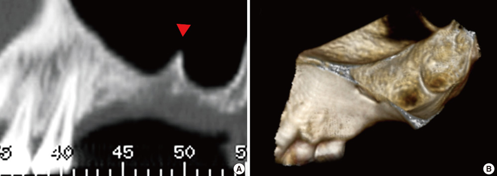
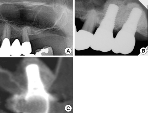
This study aimed to evaluate the effectiveness of the modified sinus floor elevation technique described hereafter as a "hybrid technique," in 11 patients with severely resorbed posterior maxillae.
METHODS
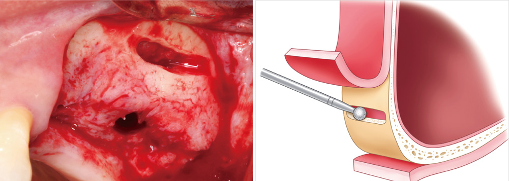
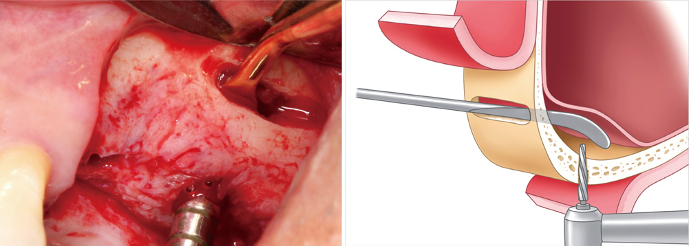
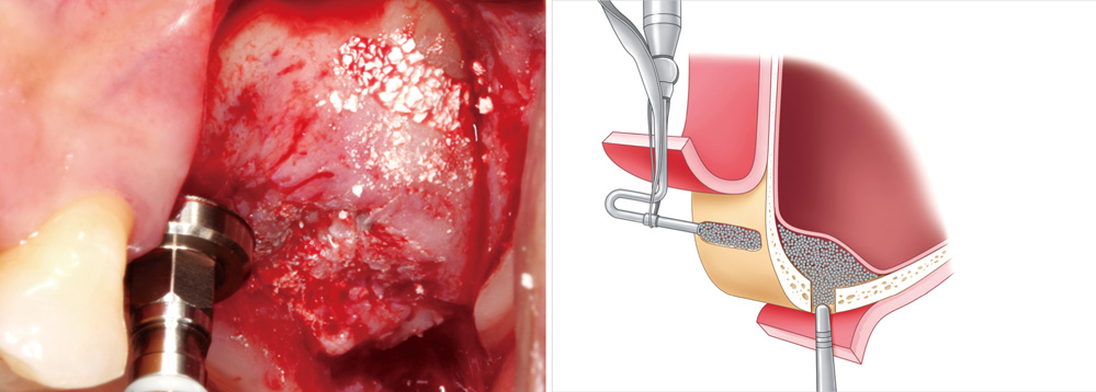
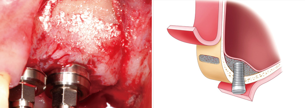
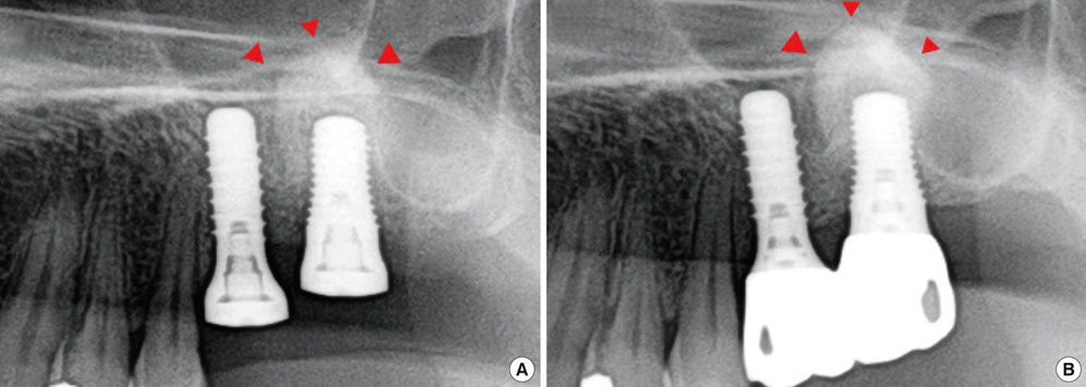
Eleven patients who received 22 implants in the maxillary premolar and molar areas by the hybrid technique were enrolled in this study. A slot-shaped osteotomy for access was prepared on the lateral wall along the lower border of the sinus floor. The Schneiderian membrane was fully reflected through the lateral slot. Following drilling with the membrane protected by a periosteal elevator, the bone was grafted. All implants were placed simultaneously with sinus augmentation. The cumulative success rate was calculated and clinical parameters were recorded. Radiographic measurements were performed.
RESULTS
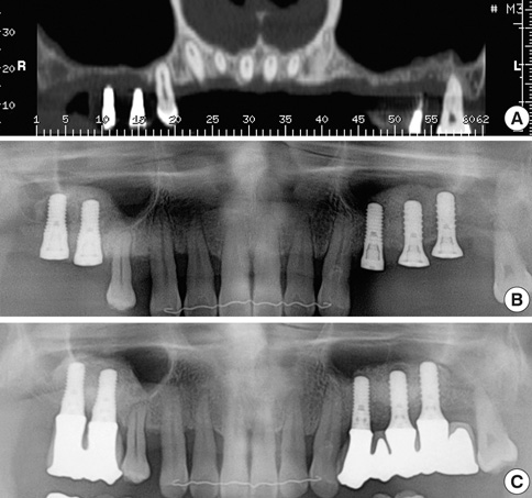
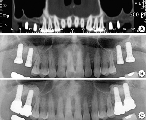
All implants were well maintained at last follow up (cumulative success rate=100%). The mean residual bone height, augmented bone height, crown-to-implant ratio, and marginal bone loss were 4.1+/-1.64 mm, 8.76+/-1.77 mm, 1.21+/-0.33 mm, and 0.34+/-0.72 mm, respectively.
CONCLUSIONS
Simultaneous implant placement with sinus augmentation by hybrid technique showed successful clinical results over a 2-year observation period and may be a reliable modality for reconstruction of a severely resorbed posterior maxilla.
Keyword
MeSH Terms
Figure
Cited by 1 articles
-
Sinus augmentation using rhBMP-2-loaded synthetic bone substitute with simultaneous implant placement in rabbits
Myung-Jae Joo, Jae-Kook Cha, Hyun-Chang Lim, Seong-Ho Choi, Ui-Won Jung
J Periodontal Implant Sci. 2017;47(2):86-95. doi: 10.5051/jpis.2017.47.2.86.
Reference
-
1. Jaffin RA, Berman CL. The excessive loss of Branemark fixtures in type IV bone: a 5-year analysis. J Periodontol. 1991. 62:2–4.
Article2. Chanavaz M. Anatomy and histophysiology of the periosteum: quantification of the periosteal blood supply to the adjacent bone with 85Sr and gamma spectrometry. J Oral Implantol. 1995. 21:214–219.3. Sharan A, Madjar D. Maxillary sinus pneumatization following extractions: a radiographic study. Int J Oral Maxillofac Implants. 2008. 23:48–56.4. Pietrokovski J, Massler M. Alveolar ridge resorption following tooth extraction. J Prosthet Dent. 1967. 17:21–27.
Article5. Boyne PJ, James RA. Grafting of the maxillary sinus floor with autogenous marrow and bone. J Oral Surg. 1980. 38:613–616.6. Geurs NC, Wang IC, Shulman LB, Jeffcoat MK. Retrospective radiographic analysis of sinus graft and implant placement procedures from the Academy of Osseointegration Consensus Conference on Sinus Grafts. Int J Periodontics Restorative Dent. 2001. 21:517–523.7. Jensen OT, Shulman LB, Block MS, Iacono VJ. Report of the Sinus Consensus Conference of 1996. Int J Oral Maxillofac Implants. 1998. 13:Suppl. 11–45.8. Pjetursson BE, Tan WC, Zwahlen M, Lang NP. A systematic review of the success of sinus floor elevation and survival of implants inserted in combination with sinus floor elevation. J Clin Periodontol. 2008. 35:216–240.
Article9. Tatum H Jr. Maxillary and sinus implant reconstructions. Dent Clin North Am. 1986. 30:207–229.10. Wallace SS, Froum SJ. Effect of maxillary sinus augmentation on the survival of endosseous dental implants. A systematic review. Ann Periodontol. 2003. 8:328–343.
Article11. Wood RM, Moore DL. Grafting of the maxillary sinus with intraorally harvested autogenous bone prior to implant placement. Int J Oral Maxillofac Implants. 1988. 3:209–214.12. Summers RB. A new concept in maxillary implant surgery: the osteotome technique. Compendium. 1994. 15:152–158.13. Pommer B, Unger E, Suto D, Hack N, Watzek G. Mechanical properties of the Schneiderian membrane in vitro. Clin Oral Implants Res. 2009. 20:633–637.14. Aimetti M, Romagnoli R, Ricci G, Massei G. Maxillary sinus elevation: the effect of macrolacerations and microlacerations of the sinus membrane as determined by endoscopy. Int J Periodontics Restorative Dent. 2001. 21:581–589.15. Becker ST, Terheyden H, Steinriede A, Behrens E, Springer I, Wiltfang J. Prospective observation of 41 perforations of the Schneiderian membrane during sinus floor elevation. Clin Oral Implants Res. 2008. 19:1285–1289.
Article16. Berengo M, Sivolella S, Majzoub Z, Cordioli G. Endoscopic evaluation of the bone-added osteotome sinus floor elevation procedure. Int J Oral Maxillofac Surg. 2004. 33:189–194.
Article17. Nkenke E, Schlegel A, Schultze-Mosgau S, Neukam FW, Wiltfang J. The endoscopically controlled osteotome sinus floor elevation: a preliminary prospective study. Int J Oral Maxillofac Implants. 2002. 17:557–566.18. Hernandez-Alfaro F, Torradeflot MM, Marti C. Prevalence and management of Schneiderian membrane perforations during sinus-lift procedures. Clin Oral Implants Res. 2008. 19:91–98.
Article19. Proussaefs P, Lozada J, Kim J, Rohrer MD. Repair of the perforated sinus membrane with a resorbable collagen membrane: a human study. Int J Oral Maxillofac Implants. 2004. 19:413–420.20. Bruschi GB, Scipioni A, Calesini G, Bruschi E. Localized management of sinus floor with simultaneous implant placement: a clinical report. Int J Oral Maxillofac Implants. 1998. 13:219–226.21. Rosen PS, Summers R, Mellado JR, Salkin LM, Shanaman RH, Marks MH, et al. The bone-added osteotome sinus floor elevation technique: multicenter retrospective report of consecutively treated patients. Int J Oral Maxillofac Implants. 1999. 14:853–858.22. Zitzmann NU, Scharer P. Sinus elevation procedures in the resorbed posterior maxilla. Comparison of the crestal and lateral approaches. Oral Surg Oral Med Oral Pathol Oral Radiol Endod. 1998. 85:8–17.23. Cutler SJ, Ederer F. Maximum utilization of the life table method in analyzing survival. J Chronic Dis. 1958. 8:699–712.
Article24. Cochran DL, Buser D, ten Bruggenkate CM, Weingart D, Taylor TM, Bernard JP. The use of reduced healing times on ITI implants with a sandblasted and acid-etched (SLA) surface: early results from clinical trials on ITI SLA implants. Clin Oral Implants Res. 2002. 13:144–153.
Article25. Friberg B. The posterior maxilla: clinical considerations and current concepts using Branemark System implants. Periodontol 2000. 2008. 47:67–78.
Article26. Vitkov L, Gellrich NC, Hannig M. Sinus floor elevation via hydraulic detachment and elevation of the Schneiderian membrane. Clin Oral Implants Res. 2005. 16:615–621.
Article27. Doud Galli SK, Lebowitz RA, Giacchi RJ, Glickman R, Jacobs JB. Chronic sinusitis complicating sinus lift surgery. Am J Rhinol. 2001. 15:181–186.
Article28. Tidwell JK, Blijdorp PA, Stoelinga PJ, Brouns JB, Hinderks F. Composite grafting of the maxillary sinus for placement of endosteal implants. A preliminary report of 48 patients. Int J Oral Maxillofac Surg. 1992. 21:204–209.
Article29. Di Girolamo M, Napolitano B, Arullani CA, Bruno E, Di Girolamo S. Paroxysmal positional vertigo as a complication of osteotome sinus floor elevation. Eur Arch Otorhinolaryngol. 2005. 262:631–633.
Article30. Penarrocha-Diago M, Rambla-Ferrer J, Perez V, Perez-Garrigues H. Benign paroxysmal vertigo secondary to placement of maxillary implants using the alveolar expansion technique with osteotomes: a study of 4 cases. Int J Oral Maxillofac Implants. 2008. 23:129–132.31. Saker M, Ogle O. Benign paroxysmal positional vertigo subsequent to sinus lift via closed technique. J Oral Maxillofac Surg. 2005. 63:1385–1387.
Article32. Betts NJ, Miloro M. Modification of the sinus lift procedure for septa in the maxillary antrum. J Oral Maxillofac Surg. 1994. 52:332–333.
Article33. Van den Bergh JP, ten Bruggenkate CM, Disch FJ, Tuinzing DB. Anatomical aspects of sinus floor elevations. Clin Oral Implants Res. 2000. 11:256–265.
Article34. Kim MJ, Jung UW, Kim CS, Kim KD, Choi SH, Kim CK, et al. Maxillary sinus septa: prevalence, height, location, and morphology. A reformatted computed tomography scan analysis. J Periodontol. 2006. 77:903–908.
Article35. Velasquez-Plata D, Hovey LR, Peach CC, Alder ME. Maxillary sinus septa: a 3-dimensional computerized tomographic scan analysis. Int J Oral Maxillofac Implants. 2002. 17:854–860.36. Pikos MA. Maxillary sinus membrane repair: report of a technique for large perforations. Implant Dent. 1999. 8:29–34.37. Small SA, Zinner ID, Panno FV, Shapiro HJ, Stein JI. Augmenting the maxillary sinus for implants: report of 27 patients. Int J Oral Maxillofac Implants. 1993. 8:523–528.38. Timmenga NM, Raghoebar GM, Boering G, van Weissenbruch R. Maxillary sinus function after sinus lifts for the insertion of dental implants. J Oral Maxillofac Surg. 1997. 55:936–939.
Article39. Cho SC, Wallace SS, Froum SJ, Tarnow DP. Influence of anatomy on Schneiderian membrane perforations during sinus elevation surgery: three-dimensional analysis. Pract Proced Aesthet Dent. 2001. 13:160–163.40. Tarnow DP, Wallace SS, Froum SJ, Rohrer MD, Cho SC. Histologic and clinical comparison of bilateral sinus floor elevations with and without barrier membrane placement in 12 patients: Part 3 of an ongoing prospective study. Int J Periodontics Restorative Dent. 2000. 20:117–125.
- Full Text Links
- Actions
-
Cited
- CITED
-
- Close
- Share
- Similar articles
-
- Clinical evaluation of Branemark Ti-Unite implant and ITI SLA implant in the post maxillary area with sinus elevation technique
- THE COMPARATIVE EVALUATION USING HATCH REAMER TECHNIQUE AND OSTEOTOME TECHNIQUE IN SINUS FLOOR ELEVATION
- Benign paroxysmal positional vertigo as a complication of sinus floor elevation
- Survival analysis of implants placed in the sinus floor elevated maxilla
- Clinical Availability of Waters' Projection in Sinus Elevation Procedures

