Imaging Findings of Localized Lymphoid Hyperplasia of the Pancreas: a Case Report
- Affiliations
-
- 1Department of Radiology, Chonnam National University Hwasun Hospital and Medical School, Hwasun-gun, Jeonnam 519-763, Korea.
- 2Department of Radiology, Chonnam National University Hospital and Medical School, Gwangju 501-757, Korea.
- 3Center for Aging and Geriatrics, Chonnam National University Hospital and Medical School, Gwangju 501-757, Korea.
- 4Department of Pathology, Chonnam National University Hospital and Medical School, Gwangju 501-757, Korea.
- KMID: 1783219
- DOI: http://doi.org/10.3348/kjr.2011.12.4.510
Abstract
- We report here on a case of localized lymphoid hyperplasia of the pancreas in a 70-year-old man which manifested as double lesions (uncinate process and tail) in the organ. The lesions were incidentally detected as hypoechoic lesions on ultrasonography and they appeared as delayed enhancing lesions on the contrast-enhanced dynamic CT and MRI. Total pancreatectomy was performed, because malignant tumor could not be excluded according to the preoperative imaging studies and the endoscopic ultrasound-guided biopsy failed. Pathology revealed localized lymphoid hyperplasia. The patient had an uneventful postoperative course. He has been alive for 18 months after surgery.
Keyword
MeSH Terms
Figure
-
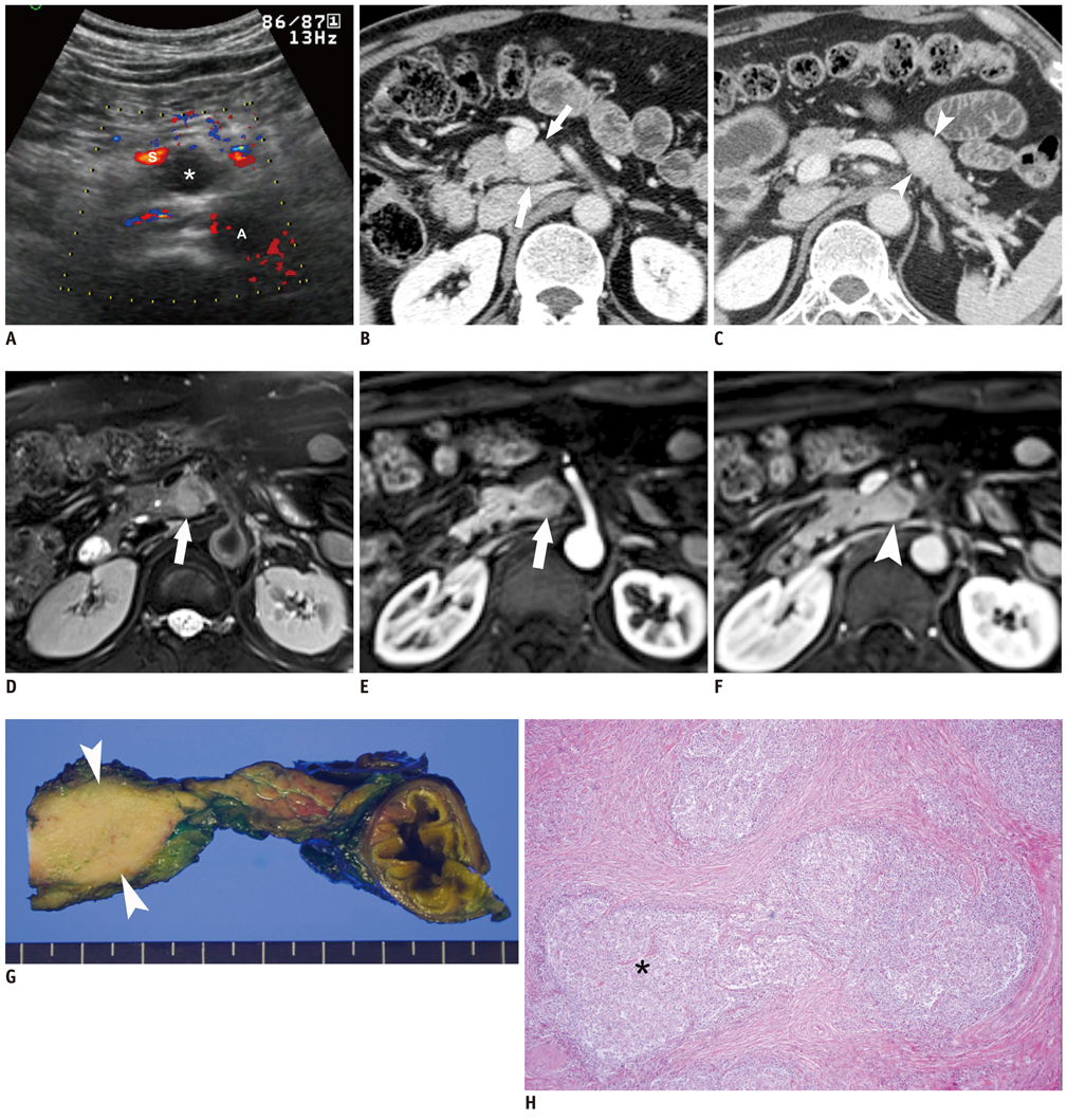
Fig. 1 Localized lymphoid hyperplasia of pancreas in 70-year-old man. A. Color Doppler ultrasonographic image shows hypoechoic mass (asterisk) in uncinate process of pancreas without discernible vascularity within mass. A = aorta, S = superior mesenteric vein. B, C. Contrast-enhanced 3-minute delayed phase CT images reveal two localized masses in uncinate process (about 2 × 1.8 cm for mass size) and tail (about 1.8 × 1.5 cm for mass size) of pancreas without peripancreatic stranding (arrows in B and arrowheads in C), and these masses show slightly more enhancement, as compared to that of pancreatic parenchyma. D. T2-weighted MR image shows localized hyperintense mass without peripancreatic stranding in uncinate process of pancreas (arrow). E, F. Gadolinium-enhanced dynamic T1-weighted images show slightly hypointense mass (arrow in E) on arterial phase image (E) in uncinate process of pancreas, and mass appears as slightly hyperintense on portal phase image (F). G. Photograph of resected specimen shows well circumscribed yellowish mass (arrowheads) in uncinate process of pancreas. H. Photomicrograph shows lymphoid follicles (asterisk) surrounded by dense collagenous tissue. Lymphoid follicles are composed of enlarged germinal centers surrounded by rims of small lymphocytes (Hematoxylin & Eosin staining, × 100). On immunohistochemical staining, neither lymphocytic infiltrate with monoclonal cell proliferation nor immunoreactivity for IgG4 was found in lesions.
Reference
-
1. Babaryka I, Thomas E. [Pancreatitis lymphomatosa (author's transl)]. Zentralbl Allg Pathol. 1981. 125:315–318.2. Hatzitheoklitos E, Buchler MW, Friess H, DiSebastiano P, Poch B, Beger HG, et al. Pseudolymphoma of the pancreas mimicking cancer. Pancreas. 1994. 9:668–670.3. Nakashiro H, Tokunaga O, Watanabe T, Ishibashi K, Kuwaki T. Localized lymphoid hyperplasia (pseudolymphoma) of the pancreas presenting with obstructive jaundice. Hum Pathol. 1991. 22:724–726.4. Machida T, Takahashi T, Itoh T, Hirayama M, Morita T, Horita S. Reactive lymphoid hyperplasia of the liver: a case report and review of literature. World J Gastroenterol. 2007. 13:5403–5407.5. Takahashi H, Sawai H, Matsuo Y, Funahashi H, Satoh M, Okada Y, et al. Reactive lymphoid hyperplasia of the liver in a patient with colon cancer: report of two cases. BMC Gastroenterol. 2006. 6:25.6. Tokunaga O, Morimatsu M, Nakashima T. Pseudolymphoma of the stomach. Nodular type. Acta Pathol Jpn. 1985. 35:227–231.7. Schwartz MS, Sherman H, Smith T, Janis R. Gastric pseudolymphoma and its relationship to malignant gastric lymphoma. Am J Gastroenterol. 1989. 84:1555–1559.8. Herwick S, Miller FH, Keppke AL. MRI of islet cell tumors of the pancreas. AJR Am J Roentgenol. 2006. 187:W472–W480.9. Kawamoto S, Siegelman SS, Hruban RH, Fishman EK. Lymphoplasmacytic sclerosing pancreatitis (autoimmune pancreatitis): evaluation with multidetector CT. Radiographics. 2008. 28:157–170.10. Merkle EM, Bender GN, Brambs HJ. Imaging findings in pancreatic lymphoma: differential aspects. AJR Am J Roentgenol. 2000. 174:671–675.11. Baek JH, Lee JM, Kim SH, Kim SJ, Lee JY, Han JK, et al. Small (<or=3 cm) solid pseudopapillary tumors of the pancreas at multiphasic multidetector CT. Radiology. 2010. 257:97–106.
- Full Text Links
-
- Actions
-
Cited
- CITED
-
- Close
- Share
-
- Similar articles
-
- Lymphoepithelial Cyst in Pancreas: a Case Report with Magnetic Resonance Imaging Findings
- A Rare Case of Multilocular Thymic Cyst with Follicular Lymphoid Hyperplasia: Radiologic and Histopathologic Features
- Radiation Treatment of Primary Orbital Lymphoid Tumors : Case Report
- Castleman's Disease in the Sigmoid Mesocolon: A Case Report
- A Case of Primary Jejunal Malignant Lymphoma Associated with Nodular Lymphoid Hyperplasia
