Pathophysiology-based Interpretation of Magnetic Resonance Imaging and Management of Cerebral Fat Embolism: Case Report and Review of Literature
- Affiliations
-
- 1Department of Neurosurgery, Kangwon National University Hospital, School of Medicine, Kangwon National University, Korea. aaapark@kangwon.ac.kr
- 2Department of Neurosurgery, In-Sung general Hospital, Korea.
- KMID: 1782996
- DOI: http://doi.org/10.13104/jksmrm.2010.14.1.69
Abstract
- Cerebral fat embolism (CFE) is a rare, albeit potentially lethal complication of long-bone fractures. All trauma patients who are initially lucid and subsequently experience mental status deterioration should undergo immediate evaluation of possible CFE. In the present case, magnetic resonance imaging (MRI) was the most sensitive technique for the diagnosis of CFE, particularly the use of diffusion-weighted images (DWI). The authors present this case to report a pathophysiology-based interpretation of the MR characteristics and treatment of CFE.
Keyword
MeSH Terms
Figure
-
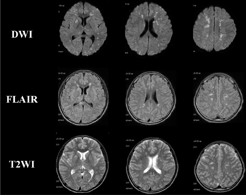
Fig. 1 MR images of the brain acquired eight hours after neurological deterioration show multiple small focal lesions in the basal ganglias, centrum semiovale, and subcortical white matter of both hemispheres. The hyperintensity lesions are most prominent on the diffusion-weighted image (DWI).
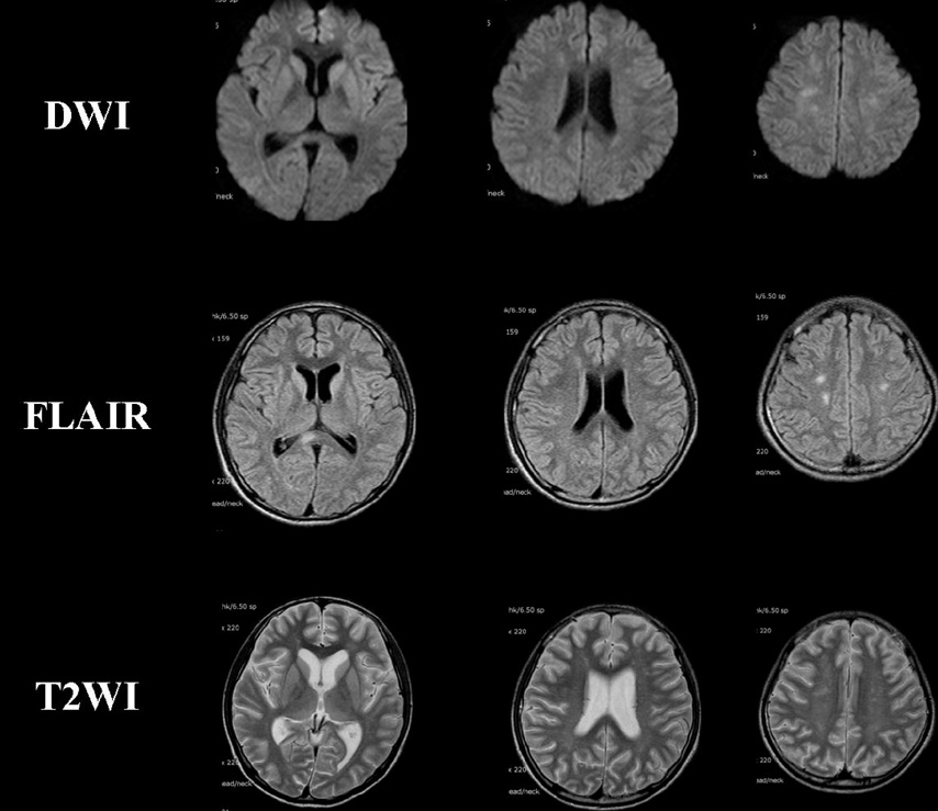
Fig. 2 Follow-up MR images of the brain performed two weeks later show a marked decrease in the size and number of hyperintensity lesion in the subcortical white matter based on DWI, FLAIR, and T2WI images.
Cited by 1 articles
-
A case of fat embolism syndrome in juvenile rheumatoid arthritis patient
Kyung Hoon Kim, Ju Kyung Lee, Young Hun Choi, Woo Sun Kim, June Dong Park, Young Yull Koh, Dong In Suh
Allergy Asthma Respir Dis. 2013;1(1):94-97. doi: 10.4168/aard.2013.1.1.94.
Reference
-
1. Johnson MJ, Lucas GL. Fat embolism syndrome. Orthopedics. 1996. 19:41–47.2. Kim HJ, Lee CH, Lee SH, et al. Early development of vasogenic edema in experimental cerebral fat embolism in cats: correlation with MRI and electron microscopic findings. Invest Radiol. 2001. 36:460–469.3. Bardana D, Rudan J, Cervenko F, Smith R. Fat embolism syndrome in a patient demonstrating only neurologic symptoms. Can J Surg. 1998. 41:398–402.4. Finlay ME, Benson MD. Case report: magnetic resonance imaging in cerebral fat embolism. Clin Radiol. 1996. 51:445–446.5. Takahashi M, Suzuki R, Osakabe Y, et al. Magnetic resonance imaging findings in cerebral fat embolism: correlation with clinical manifestations. J Trauma. 1999. 46:324–327.6. Sevitt S. The significance and pathology of fat embolism. Ann Clin Res. 1977. 9:173–180.7. Byrick RJ, Mullen JB, Mazer CD, Guest CB. Tranpulmonary systemic fat embolism: studies in mongrel dogs after cemented arthroplasty. Am J Respir Crit Care Med. 1994. 150:1416–1422.8. Beers GJ, Nichols GR, Willing SJ. CT demonstration of fat embolism associated hemorrhage in the anterior commissure. AJNR Am J Neuroradiol. 1988. 9:212–213.9. Simon AD, Ulmer JL, Strottmann JM. Contrast-enhanced MR imaging of cerebral fat embolism: case report and review of the literature. AJNR Am J Neuroradiol. 2003. 24(1):97–101.10. Lindeque BG, Schoeman H, Dommisse G, Boeyens MC, Vlok AL. Fat embolism and the fat embolism syndrome. J Bone Joint Surg Br. 1987. 69:128–131.
- Full Text Links
-
- Actions
-
Cited
- CITED
-
- Close
- Share
-
- Similar articles
-
- Gradient-Echo MRI in Defining the Severity of Cerebral Fat Embolism
- Cerebral Air Embolism: a Case Report with an Emphasis of its Pathophysiology and MRI Findings
- Cerebral Fat Embolism after Traumatic Multiple Fracture: A Case Report
- Cerebral Fat Embolism with Multiple Rib and Thoracic Spinal Fractures
- Cerebral Fat Embolism That Was Initially Negative on DiffusionWeighted Magnetic Resonance Imaging
