A Case of IgA kappa Light Chain Deposition Disease and Combined Adult Fanconi Syndrome with Auer rod-like Intracytoplasmic Inclusions in Plasma Cells and Proximal Renal Tubular Cells
- Affiliations
-
- 1Department of Laboratory Medicine, The Catholic University of Korea College of Medicine, Seoul, Korea. jmkahng@catholic.ac.kr
- 2Department of Hospital Pathology, The Catholic University of Korea College of Medicine, Seoul, Korea.
- 3Department of Internal Medicine, The Catholic University of Korea College of Medicine, Seoul, Korea.
- KMID: 1781492
- DOI: http://doi.org/10.3343/kjlm.2007.27.4.248
Abstract
- We report a case of IgA kappa light chain deposition disease and combined adult Fanconi syndrome with Auer rod-like intracytoplasmic inclusions in plasma cells and proximal renal tubular cells in a 54-yr-old female. Cytochemical stainings revealed a strong acid phosphatase activity of the inclusions and weak periodic acid-Schiff positivity, whereas the reactions for peroxidase and alpha-naphthyl acetate esterase were negative. An immunostaining verified IgA-kappa inside the plasma cells. Kidney biopsy revealed Bence Jones cast nephropathy with kappa light chain positivity, and Congo red staining was negative. Electron microscopy showed needle-shaped crystals located in tubular epithelial cells.
MeSH Terms
Figure
-
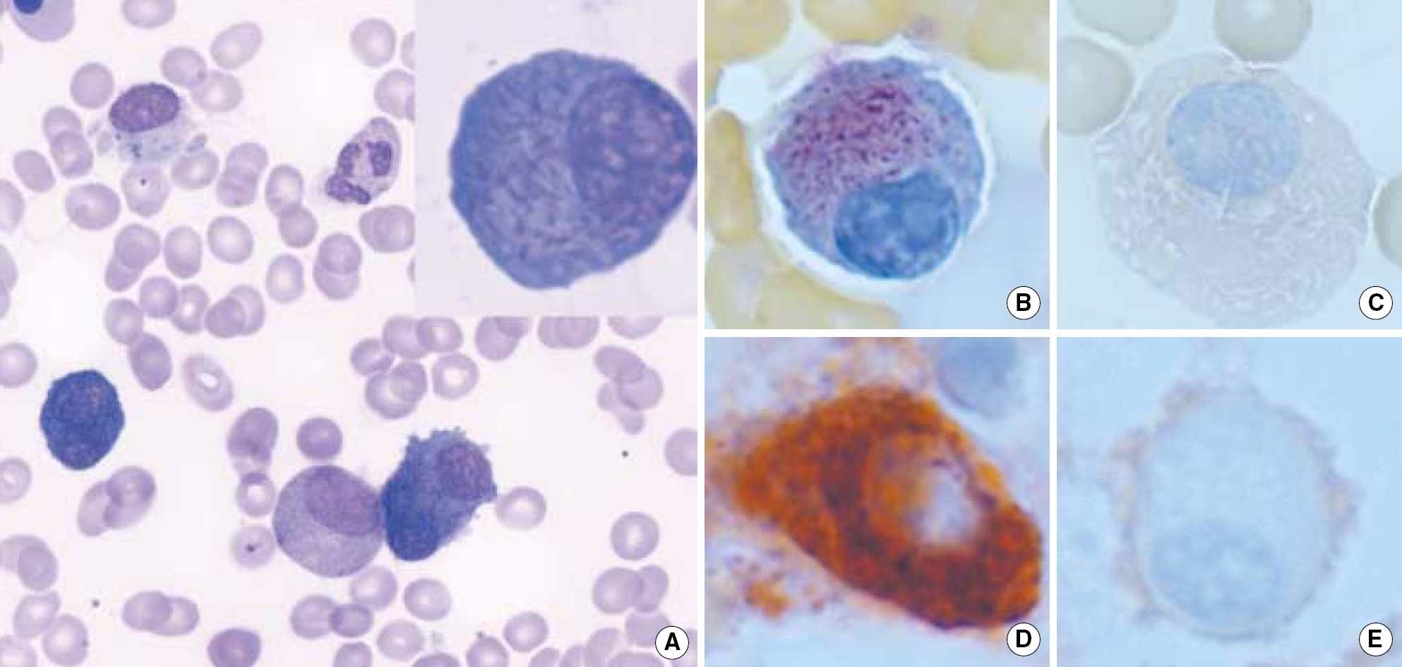
Fig. 1. Bone marrow plasma cell morphology and special stain. (A) Atypical plasma cells with spindle-shaped, Auer rod-like intracytoplasmic inclusions (Wright stain, ×400/window ×1,000). (B) Strong acid phosphatase activity of the inclusions. Note the lack of staining in surrounding cytoplasm (acid phosphatase, ×1,000). (C) Negative in non-specific esterase stain (Non-specific esterase, ×1,000). (D) and (E) Immunohistochemical stain showing kappa-positivity (D) and lambda-negativity (E) (×1,000).
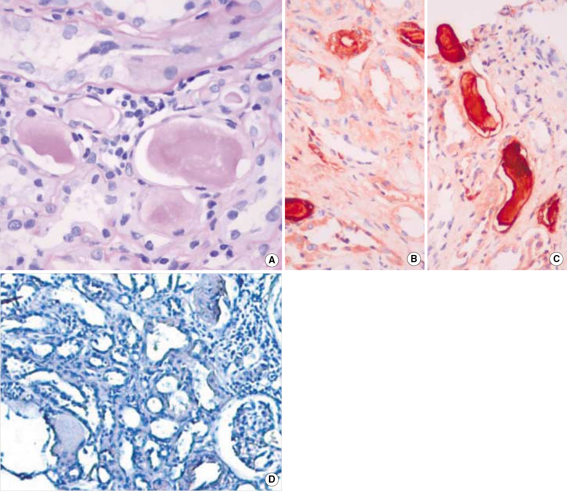
Fig. 2. The kidney biopsy revealed Bence Jones cast nephropathy with kappa light chain positivity. (A) H-E stain, ×100. (B) Immunohistochemical stain showing IgA positivity (×1,000) and (C) kappa positivity (×1,000). (D) Congo red stain showing negativity (×100).

Fig. 3. Ultrastructure of tubular epithelial cells in a case of IgA myeloma kidney. The figure shows needle-shaped crystals located in tubular epithelial cells (TEM) (A) ×2,000 & (B) ×40,000.
Reference
-
References
1. Jaffe ES, Harris NL, editors. World Health Organization classification of tumours. Pathology and genetics of tumours of haematopoietic and lymphoid tissue. France: IARC Press, Lyon;2001. p. 150–1. p. 152–4.2. Castoldi G, Piva N, Tomasi P. Multiple myeloma with Auer-rodlike inclusions. Haematologica. 1999; 84:859–60.3. Metzgeroth G, Back W, Maywald O, Schatz M, Willer A, Hehlmann R, et al. Auer rod-like inclusions in multiple myeloma. Ann Hematol. 2003; 82:57–60.
Article4. Raman SB, Van Slyck EJ. Nature of intracytoplasmic crystalline inclusions in myeloma cells (morphologic, cytochemical, ultrastructural, and immunofluorescent studies). Am J Clin Pathol. 1983; 80:224–8.
Article5. Casciaro S, Clavio M, Boccaccio P. Unusual intracellular and extracellular crystal inclusions in light chain multiple myeloma. Haematologica. 1999; 84:1046–7.6. VM, Meenu Bal M, Varma N, Sakhuja V. Auer rod-like inclusions in immunoglobulin a multiple myeloma. Arch Pathol Lab Med. 2005; 129:706–7.7. Grossniklaus HE, Stulting RD, L'Hernault N. Corneal and conjunctival crystals in paraproteinemia. Hum Pathol. 1990; 21:1181–3.
Article8. Messiaen T, Deret S, Mougenot B, Bridoux F, Dequiedt P, Dion JJ, et al. Adult Fanconi syndrome secondary to light chain gammopathy. Clinicopathologic heterogeneity and unusual features in 11 patients. Medicine. 2000; 79:135–54.9. Jung JY, Park CJ, Cho HC, Park YS. Intranuclear inclusion in IgG-lambda multiple myeloma. Korean J Clin Pathol. 1996; 16:139–42.10. Ko GA, Park CJ, Cho HC. A case of intracytoplasmic crystalline inclusions in multiple myeloma with Fanconi syndrome. Korean J Clin Pathol. 1996; 16:143–6.11. Greipp PR, Leong T, Bennett JM, Gaillard JP, Klein B, Stewart JA, et al. Plasmablastic morphology–an independent prognostic factor with clinical and laboratory correlates: Eastern Cooperative Oncology Group (ECOG) myeloma trial E9486 report by the ECOG Myeloma Laboratory Group. Blood. 1998; 91:2501–7.
Article12. Gardais J, Genevieve F, Foussard C, Delisle V, Zandecki M. Is there any significance for intracellular crystals in plasma cells from patients with monoclonal gammopathies? Eur J Haematol. 2001; 67:119–22.
Article13. Lin J, Markowitz GS, Valeri AM, Kambham N, Sherman WH, Appel GB, et al. Renal monoclonal immunoglobulin deposition disease: the disease spectrum. J Am Soc Nephrol. 2001; 12:1482–92.
Article14. Pozzi C, D'Amico M, Fogazzi GB, Curioni S, Ferrario F, Pasquali S, et al. Light chain deposition disease with renal involvement: clinical characteristics and prognostic factors. Am J Kidney Dis. 2003; 42:1154–63.
Article15. Lacy MQ, Gertz MA. Acquired Fanconi's syndrome associated with monoclonal gammopathies. Hematol Oncol Clin North Am. 1999; 13:1273–80.
Article16. Lajoie G, Leung R, Bargman JM. Clinical, biochemical, and pathological features in a patient with plasma cell dyscrasia and Fanconi syndrome. Ultrastruct Pathol. 2000; 24:221–6.
Article17. Gu X, Barrios R, Cartwright J, Font RL, Truong L, Herrera GA. Light chain crystal deposition as a manifestation of plasma cell dyscrasias: the role of immunoelectron microscopy. Hum Pathol. 2003; 34:270–7.
Article18. Asplin JR, Coe FL. Tubular disorders. Kasper DL, Braunwald E, Fauci AS, Hauser SL, Longo DL, Jameson JL, editors. Harrison's Principles of Internal Medicine. 16th ed.New York: McGraw-Hill;2004. p. 1694–702.19. Deret S, Denoroy L, Lamarine M, Vidal R, Mougenot B, Frangione B, et al. Kappa light chain-associated Fanconi's syndrome molecular analysis of monoclonal immunoglobulin light chains from patients with and without intracellular crystals. Protein Eng. 1999; 12:363–9.20. Aucouturier P, Bauwens M, Khamlichi AA, Denoroy L, Spinelli S, Touchard G, et al. Monoclonal IgL chain and L chain V domain gragment crystallization in myeloma-associated Fanconi's syndrome. J Immunol. 1993; 150:3561–8.
- Full Text Links
-
- Actions
-
Cited
- CITED
-
- Close
- Share
-
- Similar articles
-
- A Case of Adult Fanconi's Syndrome with Glomerular Podocyte Foot Process Effacement and osteomalacia Induced by k Light Chain Disease
- Plasma cells with Auer rod-like inclusions in a patient with myeloma
- Auer rod-like crystal inclusions in plasma cells of multiple myeloma
- Crystalline podocytopathy and tubulopathy without overt glomerular proteinuria in a patient with multiple myeloma
- A case of Fanconi syndrome accompanied by crystal depositions in tubular cells in a patient with multiple myeloma
