Yonsei Med J.
2010 Jan;51(1):121-127. 10.3349/ymj.2010.51.1.121.
Evaluation of Three Different Methods to Establish Animal Models of Acanthamoeba Keratitis
- Affiliations
-
- 1Department of Ophthalmology, Qilu Hospital, Shandong University, Jinan, China. xywu8868@163.com
- KMID: 1779617
- DOI: http://doi.org/10.3349/ymj.2010.51.1.121
Abstract
- PURPOSE
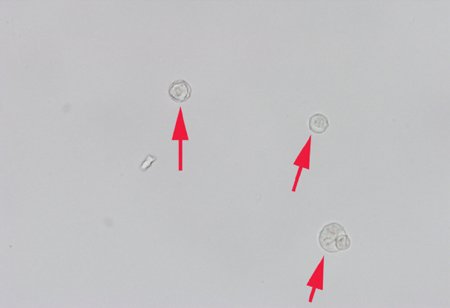
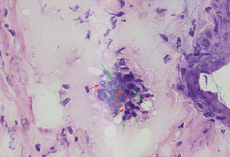
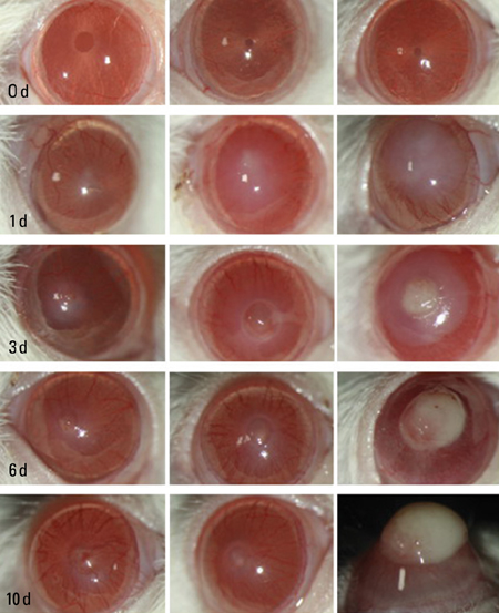
To produce animal models of Acanthamoeba keratitis and to evaluate the advantages and adaptation range of each of the three methods employed. MATERIALS AND METHODS: Mice and Wistar rats in three groups of 15 rats and 15 mice each were used to establish the models. Right corneas in group A were scratched and challenged with Acanthamoeba. Those in group B were scratched and covered with contact lenses incubated with Acanthamoeba. Those in group C received an intrastromal injection of Acanthamoeba. Five rats and 5 mice in each group were used for histopathological investigations and the other 10 in each group were used for clinical evaluation. The models were evaluated by slit lamp examination, microscopic examination and culture of corneal scrapings, HE staining of corneal sections, and pathological scoring of the infections. RESULTS: Four rats and 6 mice in group A, 7 rats and 8 mice in group B, and 10 rats and 10 mice in group C developed typical Acanthamoeba keratitis. CONCLUSION: Corneal scratching alone has the lowest infection rate, while scratching and then covering with contaminated contact lenses has a moderate rate of infection and most closely mimics what happens in most human infections. Intrastromal injection of Acanthamoeba gives a much higher infection rate and more severe Acanthamoeba keratitis.
Keyword
MeSH Terms
Figure
Reference
-
1. Jones DB, Visvesvara GS, Robinson NM. Acanthamoeba polyphaga keratitis and Acanthamoeba uveitis associated with fatal meningoencephaliti. Trans Ophthalmol Soc U K. 1975. 95:221–232.2. Seal DV, Beattie TK, Tomlinson A, Fan D, Wong E. Acanthamoeba keratitis. Br J Ophthalmol. 2003. 87:516–517.3. Wilhelmus KR, Jones DB, Matoba AY, Hamill MB, Pflugfelder SC, Weikert MP. Bilateral Acanthamoeba keratitis. Am J Ophthalmol. 2008. 145:193–197.
Article4. Gooi P, Lee-Wing M, Brownstein S, El-Defrawy S, Jackson WB, Mintsioulis G. Acanthamoeba keratitis: persistent organisms without inflammation after 1 year of topical chlorhexidine. Cornea. 2008. 27:246–248.5. Sotelo-Avila C, Taylor FM, Ewing CW. Clinical-pathological conference. Primary amebic meningoencephalitis in a healthy 7-year-old boy. J Pediatr. 1974. 85:131–136.6. Heffler KF, Eckhardt TJ, Reboli AC, Stieritz D. Acanthamoeba endophthalmitis in acquired immunodeficiency syndrome. Am J Ophthalmol. 1996. 122:584–586.
Article7. Radford CF, Bacon AS, Dart JK, Minassian DC. Risk factors for acanthamoeba keratitis in contact lens users: a case-control study. BMJ. 1995. 310:1567–1570.
Article8. Bharathi JM, Srinivasan M, Ramakrishnan R, Meenakshi R, Padmavathy S, Lalitha PN. A study of the spectrum of Acanthamoeba keratitis: a three-year study at a tertiary eye care referral center in South India. Indian J Ophthalmol. 2007. 55:37–42.
Article9. Zhang W, Pan Z, Wang ZQ, Jin XY, Luo SY, Zou Y, et al. The variance of pathogenic organisms of purulent ulcerative keratitis. Zhonghua Yan Ke Za Zhi. 2002. 38:8–12.10. Polat ZA, Ozcelik S, Vural A, Yildiz E, Cetin A. Clinical and histologic evaluations of experimental Acanthamoeba keratitis. Parasitol Res. 2007. 101:1621–1625.11. Van Klink F, Leher H, Jager MJ, Alizadeh H, Taylor W, Niederkorn JY. Systemic immune response to Acanthamoeba keratitis in the Chinese hamster. Ocul Immunol Inflamm. 1997. 5:235–244.
Article12. Alizadeh H, He Y, McCulley JP, Ma D, Stewart GL, Via M, et al. Successful immunization against Acanthamoeba keratitis in a pig model. Cornea. 1995. 14:180–186.
Article13. Awwad ST, Petroll WM, McCulley JP, Cavanagh HD. Updates in Acanthamoeba keratitis. Eye Contact Lens. 2007. 33:1–8.
Article14. Vural A, Polat ZA, Topalkara A, Toker MI, Erdogan H, Arici MK, et al. The effect of propolis in experimental Acanthamoeba Keratitis. Clin Experiment Ophthalmol. 2007. 35:749–754.
Article15. Said NA, Shoeir AT, Panjwani N, Garate M, Cao Z. Local and systemic humoral immune response during acute and chronic Acanthamoeba keratitis in rabbits. Curr Eye Res. 2004. 29:429–439.
Article16. Alizadeh H, Neelam S, Niederkorn JY. Effect of immunization with the mannose-induced Acanthamoeba protein and Acanthamoeba plasminogen activator in mitigating Acanthamoeba keratitis. Invest Ophthalmol Vis Sci. 2007. 48:5597–5604.
Article
- Full Text Links
- Actions
-
Cited
- CITED
-
- Close
- Share
- Similar articles
-
- In vivo tandem scanning confocal microscopy in acanthamoeba keratitis
- Molecular detection and characterization of Acanthamoeba infection in dogs and its association with keratitis in Korea
- The Role of Proteinase in Acanthamoeba keratitis and the Effect of Amniotic Membrane as a Proteinase Inhibitor
- Comparison of Usefulness of Laboratory Diagnosis in Ancanthamoeba Keratitis
- Two Cases of Corneal Toxicity in Acanthamoeba Keratitis by Combined Topical Anti-Acanthamoeba Keratitis Eye Solution


