Korean J Radiol.
2009 Jun;10(3):313-318. 10.3348/kjr.2009.10.3.313.
Foreign Body Granulomas Simulating Recurrent Tumors in Patients Following Colorectal Surgery for Carcinoma: a Report of Two Cases
- Affiliations
-
- 1Department of Radiology, Cheonan Hospital, Soonchunhyang University, Choongnam 330-720, Korea. rad2000@hanmail.net
- 2Department of Surgery, Cheonan Hospital, Soonchunhyang University, Choongnam 330-720, Korea.
- 3Department of Pathology, Cheonan Hospital, Soonchunhyang University, Choongnam 330-720, Korea.
- KMID: 1779461
- DOI: http://doi.org/10.3348/kjr.2009.10.3.313
Abstract
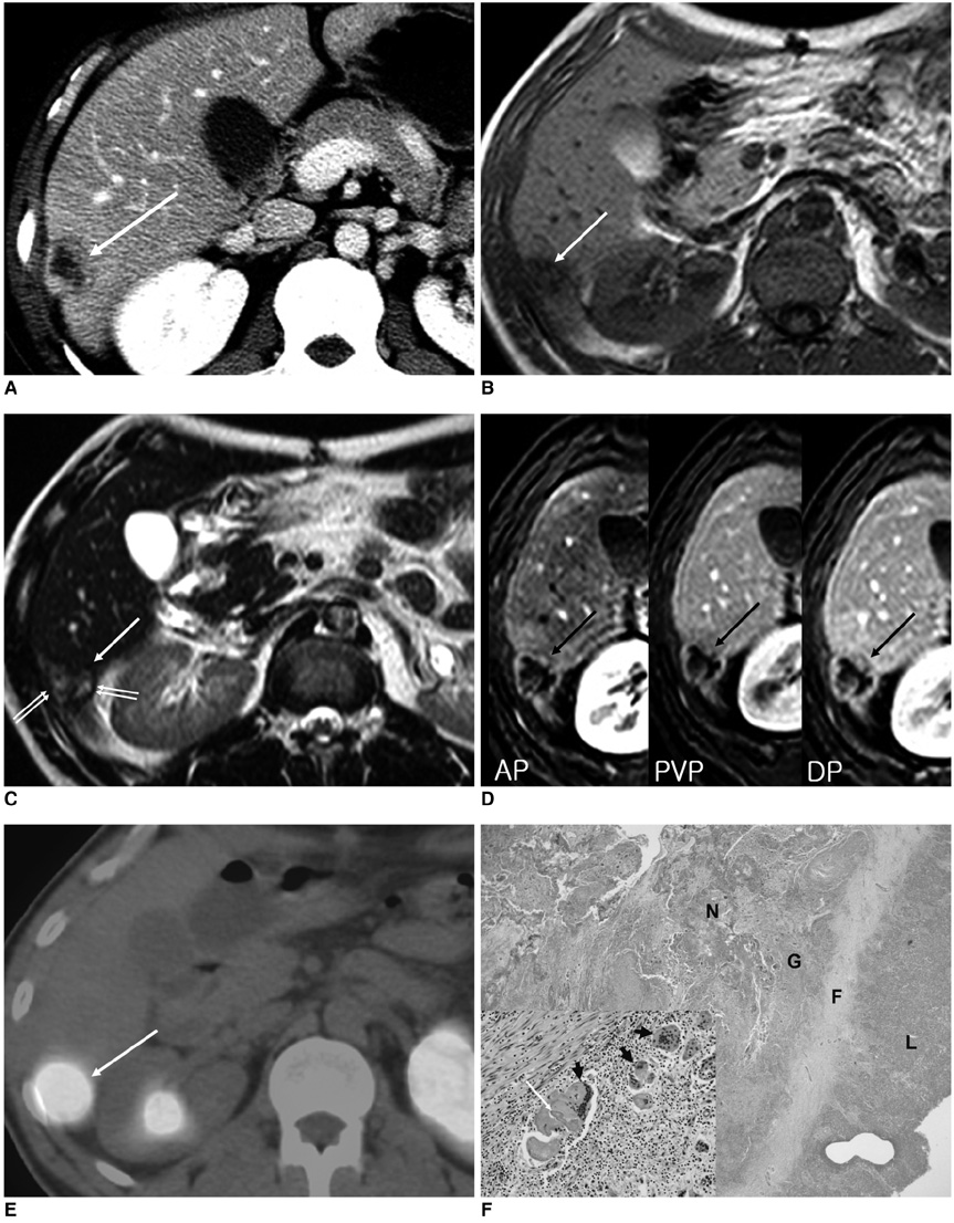
- We report here two cases of foreign body granulomas that arose from the pelvic wall and liver, respectively, and simulated recurrent colorectal carcinomas in patients with a history of surgery. On contrast-enhanced CT and MR images, a pelvic wall mass appeared as a well-enhancing mass that had invaded the distal ureter, resulting in the development of hydronephrosis. In addition, a liver mass had a hypointense rim that corresponded to the fibrous wall on a T2-weighted MR image, and showed persistent peripheral enhancement that corresponded to the granulation tissues and fibrous wall on dynamic MR images. These lesions also displayed very intense homogeneous FDG uptake on PET/CT.
Keyword
MeSH Terms
-
Adult
Aged
Colorectal Neoplasms/pathology/*surgery
Contrast Media/diagnostic use
Diagnosis, Differential
Fluorodeoxyglucose F18/diagnostic use
Granuloma, Foreign-Body/complications/*diagnosis
Humans
Hydronephrosis/etiology
Image Enhancement/methods
Liver/pathology/radionuclide imaging
Liver Neoplasms/*diagnosis/secondary
Magnetic Resonance Imaging
Male
Pelvic Neoplasms/*diagnosis/secondary
Pelvis/pathology/radiography
Positron-Emission Tomography
Radiopharmaceuticals/diagnostic use
Tomography, X-Ray Computed
Figure
Cited by 1 articles
-
Bile Granuloma Mimicking Peritoneal Seeding: A Case Report
Hasong Jeong, Hye Won Lee, Hye Ra Jung, Ilseon Hwang, Sun Young Kwon, Yu Na Kang, Sang Pyo Kim, Misun Choe
J Pathol Transl Med. 2018;52(5):339-343. doi: 10.4132/jptm.2018.06.02.
Reference
-
1. Horton KM, Abrams RA, Fishman EK. Spiral CT of colon cancer: imaging features and role in management. Radiographics. 2000. 20:419–430.2. Desch CE, Benson AB 3rd, Somerfield MR, Flynn PJ, Krause C, Loprinzi CL, et al. Colorectal cancer surveillance: 2005 update of an American Society of Clinical Oncology practice guideline. J Clin Oncol. 2005. 23:8512–8519.3. Lim JW, Tang CL, Keng GH. False positive F-18 fluorodeoxyglucose combined PET/CT scans from suture granuloma and chronic inflammation: report of two cases and review of literature. Ann Acad Med Singapore. 2005. 34:457–460.4. Poyanli A, Bilge O, Kapran Y, Güven K. Case report: foreign body granuloma mimicking liver metastasis. Br J Radiol. 2005. 78:752–754.5. Chung YE, Kim EK, Kim MJ, Yun M, Hong SW. Suture granuloma mimicking recurrent thyroid carcinoma on ultrasonography. Yonsei Med J. 2006. 47:748–751.6. Kim YK, Park HS. Foreign body granuloma of activated charcoal. Abdom Imaging. 2008. 33:94–97.7. Yeretsian RA, Blodgett TM, Branstetter BF 4th, Boberts MM, Meltzer CC. Teflon-induced granuloma: a false-positive finding with PET resolved with combined PET and CT. AJNR Am J Neuroradiol. 2003. 24:1164–1166.8. Ghersin E, Keidar Z, Brook OR, Amendola MA, Engel A. A new pitfall on abdominal PET/CT: a retained surgical sponge. J Comput Assist Tomogr. 2004. 28:839–841.9. Carroll KM, Sairam K, Olliff SP, Wallace DM. Case report: paravesical suture granuloma resembling bladder carcinoma on CT scanning. Br J Radiol. 1996. 69:476–478.10. Kise H, Shibahara T, Hayashi N, Arima K, Yanagawa M, Kawamura J. Paravesical granuloma after inguinal herniorrhaphy. Case report and review of the literature. Urol Int. 1999. 62:220–222.11. Nakajo M, Jinnouchi S, Tateno R, Nakajo M. 18F-FDG PET/CT findings of a right subphrenic foreign-body granuloma. Ann Nucl Med. 2006. 20:553–556.12. Outwater E, Tomaszewski JE, Daly JM, Kressel HY. Hepatic colorectal metastases: correlation of MR imaging and pathologic appearance. Radiology. 1991. 180:327–332.13. Danet IM, Semelka RC, Leonardou P, Braga L, Vaidean G, Woosley JT, et al. Spectrum of MRI appearances of untreated metastases of the liver. AJR Am J Roentgenol. 2003. 181:809–817.14. Metser U, Miller E, Lerman H, Even-Sapir E. Benign nonphysiologic lesions with increased 18F-FDG uptake on PET/CT: characterization and incidence. AJR Am J Roentgenol. 2007. 189:1203–1210.
- Full Text Links
- Actions
-
Cited
- CITED
-
- Close
- Share
- Similar articles
-
- Foreign Body Granulomas after the Use of Dermal Fillers: Pathophysiology, Clinical Appearance, Histologic Features, and Treatment
- Clinical Observation on Sperm Granulomas after Vasectomy
- Colorectal Foreign Bodies: Six Cases Report and Review of the Literature
- An Intradiscal Granuloma Due to a Retained Wooden Foreign Body
- Minimally invasive removal of facial foreign body granulomas


