Primary Mucinous Adenocarcinoma of a Seminal Vesicle Cyst Associated with Ectopic Ureter and Ipsilateral Renal Agenesis: a Case Report
- Affiliations
-
- 1Department of Radiology, Ilsan Paik Hospital, Inje University School of Medicine, Gyeonggi, Korea. seojwrad@ilsanpaik.ac.kr
- KMID: 1779445
- DOI: http://doi.org/10.3348/kjr.2007.8.3.258
Abstract
- Primary adenocarcinoma of the seminal vesicles is a rare neoplasm. Congenital seminal vesicle cysts are commonly associated with unilateral renal agenesis or dysgenesis. To the best of our knowledge, mucinous adenocarcinoma of the seminal vesicle cyst that's associated with an ectopic ureter opening into the seminal vesicle and ipsilateral renal agenesis has not been described in the radiological literature. We report here on the radiological findings of a primary adenocarcinoma of a seminal vesicle cyst in this condition.
MeSH Terms
Figure
-
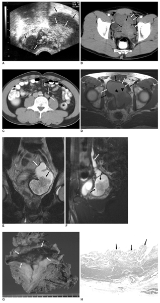
Fig. 1 A 41-year-old man with adenocarcinoma of the seminal vesicle cyst associated with an ectopic ureter opening into the seminal vesicle and also ipsilateral renal agenesis. A. Transrectal ultrasonography shows a hyperechoic, intraluminal protruding papillary mass (white arrows) in the cystic change of the left seminal vesicle (black arrows). B. On the contrast-enhanced pelvic CT scan, the papillary solid mass (black arrows) is seen to originate from the wall of the left seminal vesicle cyst (white arrows), and this mass is mildly enhanced. C. A small, abnormal soft tissue density (white arrows) is noted in the aorta's left lateral aspect, suggesting an atrophic or dysgenetic left kidney on the contrast-enhanced abdominal CT at the level of the L4 vertebra. D. An axial T1-weighted MR image shows a large, dilated seminal vesicle (white arrows) with fluid content that has high signal intensity. The cyst contains a low signal intensity papillary mass (arrowheads). The urinary bladder (black arrows) is also noted. E, F. The T2-weighted coronal (E) and sagittal (F) MR images disclose a low signal intensity papillary mass (black arrows) in the left seminal vesicle cyst and a dilated left ectopic ureter (white arrows) draining into the left seminal vesicle. G. The gross specimen shows the internal surface of the seminal vesicle cyst with a residual papillary mass (arrows). The main mass is removed. H. A photomicrograph shows a papillary configuration (black arrows) covered with carcinoma cells without muscular invasion (white arrows) (Hematoxylin & Eosin staining, × 100).
Cited by 2 articles
-
The Role of Imaging in the Diagnosis of Recurrence of Primary Seminal Vesicle Adenocarcinoma
Martina Sollini, Monica Silvotti, Massimiliano Casali, Franco Giovanardi, Alvise Zadro, Armando Froio, Paola Anna Erba, Annibale Versari
World J Mens Health. 2014;32(1):61-65. doi: 10.5534/wjmh.2014.32.1.61.Squamous Cell Carcinoma of the Seminal Vesicle from Zinner Syndrome: A Case Report and Review of Literature
Younghoon Kim, Hae Woon Baek, Eunoh Choi, Kyung Chul Moon
J Pathol Transl Med. 2015;49(1):85-88. doi: 10.4132/jptm.2014.10.28.
Reference
-
1. van den Ouden D, Blom JH, Bangma C, de Spiegeleer AH. Diagnosis and management of seminal vesicle cysts associated with ipsilateral renal agenesis: a pooled analysis of 52 cases. Eur Urol. 1998. 33:433–440.2. Schwartz ML, Kenney PJ, Bueschen AJ. Computed tomographic diagnosis of ectopic ureter with seminal vesicle cyst. Urology. 1988. 31:55–56.3. Walsh PC, Retik AB, Vaughan ED, Wein AJ. Campbell's urology. 1998. 7th ed. Philadelphia: Saunders;3299–3315.4. Levisay GL, Holder J, Weigel JW. Ureteral ectopia associated with seminal vesicle cyst and ipsilateral renal agenesis. Radiology. 1975. 114:575–576.5. Matsuki M, Matsuo M, Kaji Y, Okada N. Ectopic ureter draining into seminal vesicle cyst: usefulness of MRI. Radiat Med. 1998. 16:309–311.6. Heaney JA, Pfister RC, Meares EM Jr. Giant cyst of the seminal vesicle with renal agenesis. AJR Am J Roentgenol. 1987. 149:139–140.7. Zaontz MR, Kass EJ. Ectopic ureter opening into seminal vesicle cyst associated with ipsilateral renal agenesis. Urology. 1987. 29:523–525.8. Okada Y, Tanaka H, Takeuchi H, Yoshida O. Papillary adenocarcinoma in a seminal vesicle cyst associated with ipsilateral renal agenesis: a case report. J Urol. 1992. 148:1543–1545.9. Narita S, Akao T, Tsuchiya N, Kumazawa T, Kakinuma H, Satoh S, et al. Transitional cell carcinoma in an ectopic ureter. Int J Urol. 2003. 10:276–277.
- Full Text Links
-
- Actions
-
Cited
- CITED
-
- Close
- Share
-
- Similar articles
-
- A Case of Seminal Vesicle Cyst associated with Ipsilateral Ectopic Ureter and Renal Agenesis
- Laparoscopic Excision of Giant Seminal Vesicle Cyst Associated with Ipsilateral Renal Agenesis
- Laparoscopic Excision of Congenital Seminal Vesicle Cyst Associated with Ipsilateral Renal Agenesis
- A case of ureteral ectopia draining into seminal vesicle
- A Case of Infected Seminal Vesicle Cyst associated with Ipsilateral Renal Agenesis
