Lung Cancer Screening with Low-Dose Helical CT in Korea: Experiences at the Samsung Medical Center
- Affiliations
-
- 1Department of Radiology, Samsung Medical Center, Sungkyunkwan University School of Medicine, Seoul, Korea. kslee@smc.samsung.co.kr
- 2Division of Pulmonary and Critical Care Medicine, Samsung Medical Center, Sungkyunkwan University School of Medicine, Seoul, Korea.
- 3Department of Medicine, Center for Health Promotion, Samsung Medical Center, Sungkyunkwan University School of Medicine, Seoul, Korea.
- KMID: 1778497
- DOI: http://doi.org/10.3346/jkms.2005.20.3.402
Abstract
- To determine overall detection rates of lung cancer by low-dose CT (LDCT) screening and to compare histopathologic and imaging differences of detected cancers between high- and low-risk groups, this study included 6,406 asymptomatic Korean adults with >or=45 yr of age who underwent LDCT for lung cancer screening. All were classified into high- (>or=20 pack-year smoking; 3,353) and low-risk (3,053; <20 pack-yr smoking and non-smokers) groups. We compared CT findings of detected cancers and detection rates between high- and low-risk. At initial CT, 35% (2,255 of 6,406) had at least one or more non-calcified nodule. Lung cancer detection rates were 0.36% (23 of 6,406). Twenty-one non-small cell lung cancers appeared as solid (n=14) or ground-glass opacity (GGO) (n=7) nodules. Cancer likelihood was higher in GGO nodules than in solid nodules (p<0.01). Fifteen of 23 cancers occurred in high-risk group and 8 in low-risk group (p=0.215). Therefore, LDCT screening help detect early stage of lung cancer in asymptomatic Korean population with detection rate of 0.36% on a population basis and may be useful for discovering early lung cancer in low-risk group as well as in high-risk group.
MeSH Terms
Figure
-
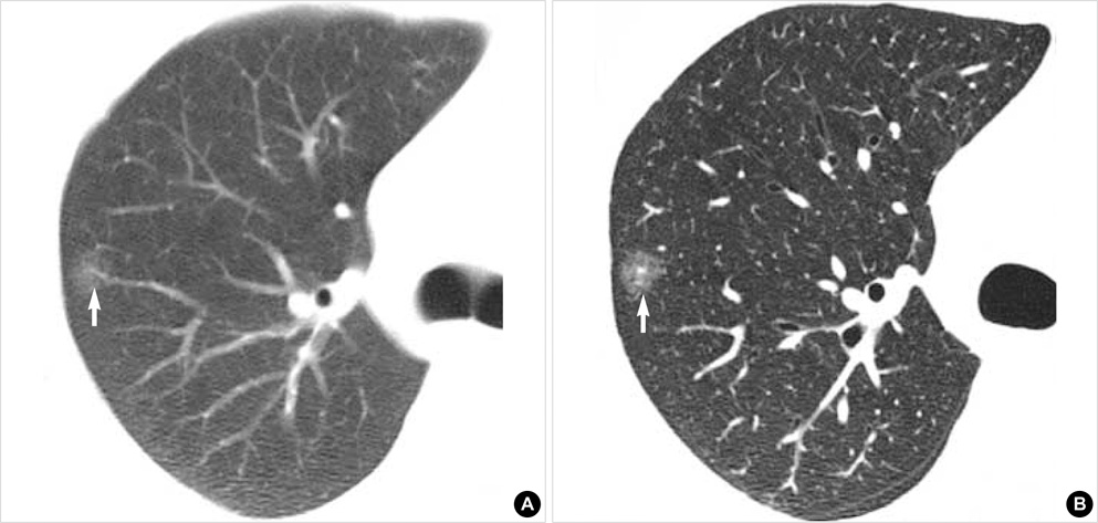
Fig. 1 A 48-yr-old man with adenocarcinoma. (A) Lung window of initial screening low-dose CT scan obtained at level of right upper lobar bronchus shows 10-mm-sized ground-glass opacity nodule (arrow) in right upper lobe. (B) Lung window of thin-section (2.5-mm thickness) CT scan obtained at similar level to A shows clearly ground-glass opacity nature of nodule (arrow). Right upper lobectomy disclosed adenocarcinoma.

Fig. 2 A 65-yr-old man with squamous cell carcinoma. (A) Lung window of initial screening low-dose CT (5-mm collimation) scan obtained at level of bronchus intermedius shows 5-mm-sized nodule (arrow) in bottom of anterior segment of right upper lobe. (B) Repeat CT scan obtained at same level to and 6 months after A shows interval increase in nodule size (arrow). Right upper lobectomy disclosed squamous cell carcinoma.
Cited by 2 articles
-
Screening for Early Detection of Cancers II
Hong Gwan Seo
J Korean Med Assoc. 2006;49(6):515-530. doi: 10.5124/jkma.2006.49.6.515.Clinical Validation of a Protein Biomarker Panel for Non-Small Cell Lung Cancer
Young Ju Jung, In-Jae Oh, Youndong Kim, Jong Ha Jung, Minkyoung Seok, Woochang Lee, Cheol Kyu Park, Jung-Hwan Lim, Young-Chul Kim, Woo-Sung Kim, Chang-Min Choi
J Korean Med Sci. 2018;33(53):. doi: 10.3346/jkms.2018.33.e342.
Reference
-
1. Greenlee RT, Murray T, Bolden S, Wingo PA. Cancer statistics, 2000. CA Cancer J Clin. 2000. 50:7–33.
Article2. Cancer statistics 2004. American Cancer Society Web site. accessed 20 July 2004. Available at: www.cancer.org/downloads/PRO/Cancer%20Statistics%202004.ppt.3. Annual Report of the Korea Central Cancer Registry. National Cancer Center Web site. accessed 20 July 2004. Available at: www.ncc.re.kr/files/cancerStat/2002_cancer_regi.ppt.4. Annual Report on the Case of Death Statistics (Based on Vital Registration). Korean National Statistical Office Web site. accessed 23 October 2004. Available at: www.nso.go.kr/newnso/upload_file/upload2/svca0300.pdf.5. Cancer facts and figures 2003. American Cancer Society Web site. accessed 20 July 2004. Available at: www.cancer.org/downloads/STT/CAFF2003PWSecured.pdf.6. Berrino R, Capocaccia R, Esteve J. Survival of cancer patients in Europe: the EUROCARE-2 study. 1999. Lyon, France: IARC Scientific Publications;1–572.7. NHS Performance Indicators National Figures: February 2002. Department of Health Web site. accessed 20 July 2004. Available at: www.performance.doh.gov.uk/nhsperformanceindicators/hlpi2002/NationalDocument.pdf.8. Flehinger BJ, Kimmel M, Melamed MR. The effect of surgical treatment on survival from early lung cancer. Implications for screening. Chest. 1992. 101:1013–1018.9. Noguchi M, Morikawa A, Kawasaki M, Matsuno Y, Yamada T, Hirohashi S, Kondo H, Shimosato Y. Small adenocarcinoma of the lung. Histologic characteristics and prognosis. Cancer. 1995. 75:2844–2852.
Article10. Frost JK, Ball WC Jr, Levin ML, Tockman MS, Baker RR, Carter D, Eggleston JC, Erozan YS, Gupta PK, Khouri NF. Early lung cancer detection: results of the initial (prevalence) radiologic and cytologic screening in the Johns Hopkins study. Am Rev Resp Dis. 1984. 130:549–554.11. Fontana RS, Sanderson DR, Taylor WF, Woolner LB, Miller WE, Muhm JR, Uhlenhopp MA. Early lung cancer detection: results of the initial (prevalence) radiologic and cytologic screening in the Mayo Clinic Study. Am Rev Resp Dis. 1984. 130:561–565.12. Melamed MR, Flehinger BJ, Zaman MB, Heelan RT, Perchick WA, Martini N. Screening for early lung cancer: results of the Memorial Sloan-Kettering study in New York. Chest. 1984. 86:44–53.13. Kubik A, Polak J. Lung cancer detection: results of a randomized prospective study in Czechoslovakia. Cancer. 1986. 57:2427–2437.
Article14. Eddy DM. Screening for lung cancer. Ann Int Med. 1989. 111:232–237.
Article15. Sone S, Li F, Yang ZG, Takashima S, Maruyama Y, Hasegawa M, Wang JC, Kawakami S, Honda T. Characteristics of small lung cancers invisible on conventional chest radiography and detected by population based screening using spiral CT. Br J Radiol. 2000. 73:137–145.
Article16. Kaneko M, Eguchi K, Ohmatsu H, Kakinuma R, Naruke T, Suemasu K, Moriyama N. Peripheral lung cancer: screening and detection with low-dose spiral CT versus radiography. Radiology. 1996. 201:798–802.
Article17. Rusinek H, Naidich DP, McGuinness G, Leitman BS, McCauley DI, Krinsky GA, Clayton K, Cohen H. Pulmonary nodule detection: low-dose versus conventional CT. Radiology. 1998. 209:243–249.
Article18. Swensen SJ, Jett JR, Sloan JA, Midthun DE, Hartman TE, Sykes AM, Aughenbaugh GL, Zink FE, Hillman SL, Noetzel GR, Marks RS, Clayton AC, Pairolero PC. Screening for lung cancer with low-dose spiral computed tomography. Am J Respir Crit Care Med. 2002. 165:508–513.
Article19. Mountain CF, Dresler CM. Regional lymph node classification for lung cancer staging. Chest. 1997. 111:1718–1723.
Article20. Green RA, Humphrey E, Close H. Alkylating agents in bronchogenic carcinoma. Am J Med. 1969. 46:516–525.
Article21. Ozono S, Miyao N, Igarashi T, Marumo K, Nakazawa H, Fukuda M, Tsushima T, Tokuda N, Kawamura J, Murai M. Tumor doubling time of renal cell carcinoma measured by CT: collaboration of Japanese Society of Renal Cancer. Jpn J Clin Oncol. 2004. 34:82–85.
Article22. Sobue T, Moriyama N, Kaneko M, Kusumoto M, Kobayashi T, Tsuchiya R, Kakinuma R, Ohmatsu H, Nagai K, Nishiyama H, Matsui E, Eguchi K. Screening for lung cancer with low-dose helical computed tomography: anti-lung cancer association project. J Clin Oncol. 2002. 20:911–920.
Article23. Sone S, Li F, Yang ZG, Honda T, Maruyama Y, Takashima S, Hasegawa M, Kawakami S, Kubo K, Haniuda M, Yamanda T. Results of three-year mass screening programme for lung cancer using mobile low-dose spiral computed tomography scanner. Br J Cancer. 2001. 84:25–32.
Article24. Henschke CI, Yankelevitz DF, Libby DM, McCauley D, Pasmantier M, Altorki NK, Smith JP, Miettinen OS. Early lung cancer action project: annual screening using single-slice helical CT. Ann N Y Acad Sci. 2001. 952:124–134.25. Swensen SJ, Jett JR, Hartman TE, Midthun DE, Sloan JA, Sykes AM, Aughenbaugh GL, Clemens MA. Lung cancer screening with CT: Mayo Clinic experience. Radiology. 2003. 226:756–761.
Article26. Diederich S, Wormanns D, Semik M, Diederich S, Wormanns D, Semik M, Thomas M, Lenzen H, Roos N, Heindel W. Screening for early lung cancer with low-dose spiral CT: prevalence in 817 asymptomatic smokers. Radiology. 2002. 222:773–781.
Article27. Nakata M, Saeki H, Takata I, Segawa Y, Mogami H, Mandai K, Eguchi K. Focal ground-glass opacity detected by low-dose helical CT. Chest. 2002. 121:1464–1467.
Article28. Kakinuma R, Ohmatsu H, Kaneko M, Kusumoto M, Yoshida J, Nagai K, Nishiwaki Y, Kobayashi T, Tsuchiya R, Nishiyama H, Matsui E, Eguchi K, Moriyama N. Progression of focal pure ground-glass opacity detected by low-dose helical computed tomography screening for lung cancer. J Comput Assist Tomogr. 2004. 28:17–23.
Article
- Full Text Links
-
- Actions
-
Cited
- CITED
-
- Close
- Share
-
- Similar articles
-
- Screening for Lung Cancer
- Korean Society of Thoracic Radiology Guideline for Lung Cancer Screening with Low-Dose CT
- Lung Cancer Screening with Low-dose Computed Tomography
- Lung cancer screening with low-dose chest computed tomography: recent radiologic advances
- Screening for Lung Cancer with Low-Dose Computed Tomography
