A Case of Granulocyte-Colony Stimulating Factor-Producing Hepatocellular Carcinoma Confirmed by Immunohistochemistry
- Affiliations
-
- 1Department of Internal Medicine, Matsumoto Medical Center, Matsumoto, Japan. joshita@shinshu-u.ac.jp
- 2Department of Internal Medicine, Division of Gastroenterology and Hepatology, Shinshu University School of Medicine, Matsumoto, Japan.
- 3Department of Laboratory Medicine, Matsumoto Medical Center, Matsumoto, Japan.
- 4Department of Surgery, Matsumoto Medical Center, Matsumoto, Japan.
- KMID: 1778047
- DOI: http://doi.org/10.3346/jkms.2010.25.3.476
Abstract
- Granulocyte-colony stimulating factor (G-CSF) is a naturally occurring glycoprotein that stimulates the proliferation and maturation of precursor cells in the bone marrow into fully differentiated neutrophils. Several reports of G-CSF-producing malignant tumors have been published, but scarcely any in the hepatobiliary system, such as in hepatocellular carcinoma (HCC). Here, we encountered a 69-yr-old man with a hepatic tumor who had received right hepatic resection. He showed leukocytosis of 25,450/microL along with elevated serum G-CSF. Histological examination of surgical samples demonstrated immunohistochemical staining for G-CSF, but not for G-CSF receptor. The patient survived without recurrence for four years, but ultimately passed away with multiple bone metastases. In light of the above, clinicians may consider G-CSF-producing HCC when encountering patients with leukocytosis and a hepatic tumor. More cases are needed to clarify the clinical picture of G-CSF-producing HCC.
MeSH Terms
-
Aged
Bone Neoplasms/secondary
Carcinoma, Hepatocellular/*metabolism/pathology
Fatal Outcome
Granulocyte Colony-Stimulating Factor/*metabolism
Humans
Liver Neoplasms/*metabolism/pathology
Male
Receptors, Granulocyte Colony-Stimulating Factor/metabolism
Receptors, Granulocyte Colony-Stimulating Factor
Granulocyte Colony-Stimulating Factor
Figure
-

Fig. 1 Computed tomography findings. (A) The tumor measuring 5 cm in diameter between the anterior inferior segment (S5) and the anterior superior segment (S8) of the liver showed hyper-enhancement (black arrow heads) in the early phase of dynamic enhanced CT accompanied with diffuse enhancement in the surrounding area (white circle). (B) It showed complete washout (black arrow heads) in the late phase with delayed hyper-enhancement in the surrounding area (white circle).
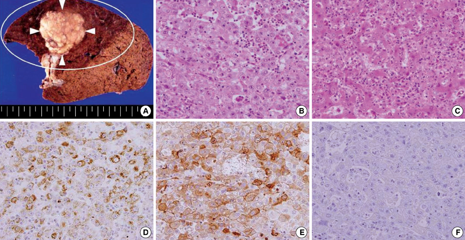
Fig. 2 Gross and microscopic findings of the tumor. (A) Cut surface of the resected liver showed an encapsulated gray-white nodule (white arrow heads) with foci of necrosis. The area adjacent to the tumor (white circle) revealed prominent congestion. Non-neoplastic liver parenchyma was not cirrhotic. (B) Microscopic findings showed atypical cells lying in sheets with marked infiltration of neutrophils and lymphocytes, which were diagnosed as a moderately differentiated hepatocellular carcinoma (H&E, ×20 magnification of the objective lens). (C) Liver parenchyma adjacent to the tumor, diffusely enhanced by contrast enhanced CT, showed prominent congestion and marked infiltration with neutrophils within the widened sinusoid (H&E, ×20 magnification of the objective lens). The other parts of liver did not present findings of chronic hepatitis or cirrhosis (not shown). (D) The tumor lesion was stained with hepatocyte paraffin 1 (Hep par 1) (×20 magnification of the objective lens). (E) Immunohistochemical examination also showed positive staining for granulocyte-colony stimulating factor (G-CSF) in the cytoplasm of atypical cells (×20 magnification of the objective lens). (F) Immunohistochemical examination showed negative staining for G-CSF receptors in the tumor cells (×20 magnification of the objective lens).
Reference
-
1. Bradley TR, Metcalf D. The growth of mouse bone marrow cells in vitro. Aust J Exp Biol Med Sci. 1966. 44:287–299.2. Robinson WA. Granulocytosis in neoplasia. Ann N Y Acad Sci. 1974. 230:212–218.
Article3. Asano S, Urabe A, Okabe T, Sato N, Kondo Y. Demonstration of granulopoietic factor(s) in the plasma of nude mice transplanted with a human lung cancer and in the tumor tissue. Blood. 1977. 49:845–852.
Article4. Lieschke GJ, Burgess AW. Granulocyte colony-stimulating factor and granulocyte-macrophage colony-stimulating factor (1). N Engl J Med. 1992. 327:28–35.5. Yamamoto S, Takashima S, Ogawa H, Kuroda T, Yamamoto M, Takeda A, Nakmaura H. Granulocyte-colony-stimulating-factor-producing hepatocellular carcinoma. J Gastroenterol. 1999. 34:640–644.
Article6. Araki K, Kishihara F, Takahashi K, Matsumata T, Shimura T, Suehiro T, Kuwano H. Hepatocellular carcinoma producing a granulocyte colony-stimulating factor: report of a resected case with a literature review. Liver Int. 2007. 27:716–721.
Article7. Shimamura K, Fujimoto J, Hata J, Akatsuka A, Ueyama Y, Watanabe T, Tamaoki N. Establishment of specific monoclonal antibodies against recombinant human granulocyte colony-stimulating factor (hG-CSF) and their application for immunoperoxidase staining of paraffin-embedded sections. J Histochem Cytochem. 1990. 38:283–286.
Article8. Higaki I, Hirohashi K, Fukushima S, Wanibuchi H, Seike N, Yamane T, Kubo S, Tanaka H, Shuto T, Yamamoto T, Kinoshita H. Renal pelvic carcinoma producing granulocyte colony-stimulating factor: report of a case. Surg Today. 2001. 31:266–268.
Article9. Segawa K, Ueno Y, Kataoka T. In vivo tumor growth enhancement by granulocyte colony-stimulating factor. Jpn J Cancer Res. 1991. 82:440–447.
Article10. Tachibana M, Miyakawa A, Tazaki H, Nakamura K, Kubo A, Hata J, Nishi T, Amano Y. Autocrine growth of transitional cell carcinoma of the bladder induced by granulocyte-colony stimulating factor. Cancer Res. 1995. 55:3438–3443.11. Baba M, Hasegawa H, Nakayabu M, Shimizu N, Suzuki S, Kamada N, Tani K. Establishment and characteristics of a gastric cancer cell line (HuGC-OOHIRA) producing high levels of G-CSF, GM-CSF, and IL-6: the presence of autocrine growth control by G-CSF. Am J Hematol. 1995. 49:207–215.
Article12. Kyo S, Kanaya T, Takakura M, Inoue M. A case of cervical cancer with aggressive tumor growth: possible autocrine growth stimulation by G-CSF and Il-6. Gynecol Oncol. 2000. 78:383–387.
Article13. Mueller MM, Herold-Mende CC, Riede D, Lange M, Steiner HH, Fusenig NE. Autocrine growth regulation by granulocyte colony-stimulating factor and granulocyte macrophage colony-stimulating factor in human gliomas with tumor progression. Am J Pathol. 1999. 155:1557–1567.
Article14. Tsuzuki H, Fujieda S, Sunaga H, Noda I, Saito H. Expression of granulocyte colony-stimulating factor receptor correlates with prognosis in oral and mesopharyngeal carcinoma. Cancer Res. 1998. 58:794–800.15. Suzuki A, Takahashi T, Okuno Y, Tsuyuoka R, Fukumoto M, Nakamura K, Imura H. IL-1 production as a regulator of G-CSF and IL-6 production in CSF-producing cell lines. Br J Cancer. 1992. 65:515–518.
Article16. Dinarello CA. Cytokines as endogenous pyrogens. J Infect Dis. 1999. 179:Suppl 2. S294–S304.
Article17. Luheshi GN. Cytokines and fever. Mechanisms and sites of action. Ann N Y Acad Sci. 1998. 856:83–89.
Article18. Castell JV, Gomez-Lechon MJ, David M, Andus T, Geiger T, Trullenque R, Fabra R, Heinrich PC. Interleukin-6 is the major regulator of acute phase protein synthesis in adult human hepatocytes. FEBS Lett. 1989. 242:237–239.
Article19. Castell JV, Gomez-Lechon MJ, David M, Fabra R, Trullenque R, Heinrich PC. Acute-phase response of human hepatocytes: regulation of acute-phase protein synthesis by interleukin-6. Hepatology. 1990. 12:1179–1186.
Article
- Full Text Links
-
- Actions
-
Cited
- CITED
-
- Close
- Share
-
- Similar articles
-
- Sweet Syndrome in a Child with Aplastic Anemia after Receiving Recombinant Granulocyte Colony-stimulating Factor
- Two cases of congenital agranulocytosis treated with recombinant human granulocyte colony-stimulating factor
- The effect of granulocyte colony-stimulating factor in chemotherapy of acute myelogenous leukemia
- The effects on the production of platelet activating factor in the cultured human endothelial cells by interleukin-6 and granulocyte macrophage colony stimulating factor
- A Metastatic Granulocyte Colony-Stimulating Factor Producing Sarcomatoid Carcinoma of the Lung Causing Jejunal Intussusception: Report of a Case
