J Korean Med Sci.
2013 May;28(5):744-749. 10.3346/jkms.2013.28.5.744.
Interobserver Variability and Accuracy of High-Definition Endoscopic Diagnosis for Gastric Intestinal Metaplasia among Experienced and Inexperienced Endoscopists
- Affiliations
-
- 1Department of Internal Medicine, Hanyang University Guri Hospital, Guri, Korea. hands@hanyang.ac.kr
- KMID: 1777563
- DOI: http://doi.org/10.3346/jkms.2013.28.5.744
Abstract
- Accurate diagnosis of gastric intestinal metaplasia is important; however, conventional endoscopy is known to be an unreliable modality for diagnosing gastric intestinal metaplasia (IM). The aims of the study were to evaluate the interobserver variation in diagnosing IM by high-definition (HD) endoscopy and the diagnostic accuracy of this modality for IM among experienced and inexperienced endoscopists. Selected 50 cases, taken with HD endoscopy, were sent for a diagnostic inquiry of gastric IM through visual inspection to five experienced and five inexperienced endoscopists. The interobserver agreement between endoscopists was evaluated to verify the diagnostic reliability of HD endoscopy in diagnosing IM, and the diagnostic accuracy, sensitivity, and specificity were evaluated for validity of HD endoscopy in diagnosing IM. Interobserver agreement among the experienced endoscopists was "poor" (kappa = 0.38) and it was also "poor" (kappa = 0.33) among the inexperienced endoscopists. The diagnostic accuracy of the experienced endoscopists was superior to that of the inexperienced endoscopists (P = 0.003). Since diagnosis through visual inspection is unreliable in the diagnosis of IM, all suspicious areas for gastric IM should be considered to be biopsied. Furthermore, endoscopic experience and education are needed to raise the diagnostic accuracy of gastric IM.
MeSH Terms
Figure
-
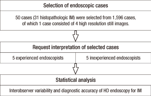
Fig. 1 Flow diagram of the process of image evaluation.
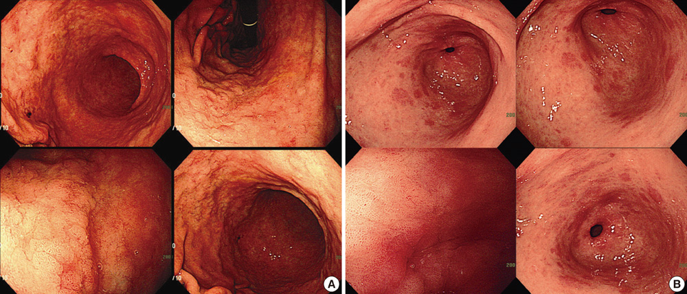
Fig. 2 Examples of a high-definition endoscopic case consisting of four images for interpretation. The close-up image shows the sharp mucosal surface of the biopsy site. The three other images include the site of biopsy from different angles and distances. (A) Nine out of 10 endoscopists agreed with the diagnosis of gastric IM on the anterior wall of lower body, and histopathology proved to be marked IM. (B) Two out of 10 endoscopists agreed with the diagnosis of gastric IM on the great curvature of antrum, and histopathology proved to be moderate IM.
Reference
-
1. Correa P. Human gastric carcinogenesis: a multistep and multifactorial process: first American Cancer Society Award Lecture on Cancer Epidemiology and Prevention. Cancer Res. 1992. 52:6735–6740.2. De Vries AC, van Grieken NC, Looman CW, Casparie MK, de Vries E, Meijer GA, Kuipers EJ. Gastric cancer risk in patients with premalignant gastric lesions: a nationwide cohort study in the Netherlands. Gastroenterology. 2008. 134:945–952.3. Correa P, Piazuelo MB, Wilson KT. Pathology of gastric intestinal metaplasia: clinical implications. Am J Gastroenterol. 2010. 105:493–498.4. Kaur G, Raj SM. A study of the concordance between endoscopic gastritis and histological gastritis in an area with a low background prevalence of Helicobacter pylori infection. Singapore Med J. 2002. 43:090–092.5. Laine L, Cohen H, Sloane R, Marin-Sorensen M, Weinstein WM. Interobserver agreement and predictive value of endoscopic findings for H. pylori and gastritis in normal volunteers. Gastrointest Endosc. 1995. 42:420–423.6. Kaminishi M, Yamaguchi H, Nomura S, Oohara T, Sakai S, Fukutomi H, Nakahara A, Kashimura H, Oda M, Kitahora T, et al. Endoscopic classification of chronic gastritis based on a pilot study by the research society for gastritis. Dig Endosc. 2002. 14:138–151.7. Areia M, Amaro P, Dinis-Ribeiro M, Cipriano MA, Marinho C, Costa-Pereira A, Lopes C, Moreira-Dias L, Romãozinho JM, Gouveia H, et al. External validation of a classification for methylene blue magnification chromoendoscopy in premalignant gastric lesions. Gastrointest Endosc. 2008. 67:1011–1018.8. Bansal A, Ulusarac O, Mathur S, Sharma P. Correlation between narrow band imaging and nonneoplastic gastric pathology: a pilot feasibility trial. Gastrointest Endosc. 2008. 67:210–216.9. Capelle LG, Haringsma J, de Vries AC, Steyerberg EW, Biermann K, van Dekken H, Kuipers EJ. Narrow band imaging for the detection of gastric intestinal metaplasia and dysplasia during surveillance endoscopy. Dig Dis Sci. 2010. 55:3442–3448.10. Guo YT, Li YQ, Yu T, Zhang TG, Zhang JN, Liu H, Liu FG, Xie XJ, Zhu Q, Zhao YA. Diagnosis of gastric intestinal metaplasia with confocal laser endomicroscopy in vivo: a prospective study. Endoscopy. 2008. 40:547–553.11. Adler A, Aschenbeck J, Yenerim T, Mayr M, Aminalai A, Drossel R, Schröder A, Scheel M, Wiedenmann B, Rösch T. Narrow-band versus whitelight high definition television endoscopic imaging for screening colonoscopy: a prospective randomized trial. Gastroenterology. 2009. 136:410–416.12. Rex DK, Helbig CC. High yields of small and flat adenomas with high-definition colonoscopes using either white light or narrow band imaging. Gastroenterology. 2007. 133:42–47.13. Curvers W, Baak L, Kiesslich R, Van Oijen A, Rabenstein T, Ragunath K, Rey JF, Scholten P, Seitz U, Ten Kate F, et al. Chromoendoscopy and narrow-band imaging compared with high-resolution magnification endoscopy in Barrett's esophagus. Gastroenterology. 2008. 134:670–679.14. Aabakken L, Rembacken B, LeMoine O, Kuznetsov K, Rey JF, Rösch T, Eisen G, Cotton P, Fujino M. Minimal standard terminology for gastrointestinal endoscopy - MST 3.0. Endoscopy. 2009. 41:727–728.15. Flahault A, Cadilhac M, Thomas G. Sample size calculation should be performed for design accuracy in diagnostic test studies. J Clin Epidemiol. 2005. 58:859–862.16. Chmura Kraemer H, Periyakoil VS, Noda A. Kappa coefficients in medical research. Stat Med. 2002. 21:2109–2129.17. McGraw KO, Wong SP. Forming inferences about some intraclass correlation coefficients. Psychological Methods. 1996. 1:30–46.18. Stokes ME, Davis CS, Koch GG. Categorical data analysis using the SAS system. 2000. 2nd ed. Cary: SAS Institute Inc..19. Van den Broek FJ, van Soest EJ, Naber AH, van Oijen AH, Mallant-Hent RCh, Böhmer CJ, Scholten P, Stokkers PC, Marsman WA, Mathus-Vliegen EM, et al. Combining autofluorescence imaging and narrow-band imaging for the differentiation of adenomas from non-neoplastic colonic polyps among experienced and non-experienced endoscopists. Am J Gastroenterol. 2009. 104:1498–1507.20. Dixon MF, Genta RM, Yardley JH, Correa P. Classification and grading of gastritis: the updated Sydney System: International Workshop on the Histopathology of Gastritis, Houston 1994. Am J Surg Pathol. 1996. 20:1161–1181.21. Sauerbruch T, Schreiber MA, Schüssler P, Permanetter W. Endoscopy in the diagnosis of gastritis: diagnostic value of endoscopic criteria in relation to histological diagnosis. Endoscopy. 1984. 16:101–104.22. Choi IJ. Gastric cancer screening and diagnosis. Korean J Gastroenterol. 2009. 54:67–76.23. Rabeneck L, Paszat LF, Saskin R. Endoscopist specialty is associated with incident colorectal cancer after a negative colonoscopy. Clin Gastroenterol Hepatol. 2010. 8:275–279.24. Baxter NN, Sutradhar R, Forbes SS, Paszat LF, Saskin R, Rabeneck L. Analysis of administrative data finds endoscopist quality measures associated with postcolonoscopy colorectal cancer. Gastroenterology. 2011. 140:65–72.25. Higashi R, Uraoka T, Kato J, Kuwaki K, Ishikawa S, Saito Y, Matsuda T, Ikematsu H, Sano Y, Suzuki S, et al. Diagnostic accuracy of narrow-band imaging and pit pattern analysis significantly improved for less-experienced endoscopists after an expanded training program. Gastrointest Endosc. 2010. 72:127–135.26. Chang CC, Hsieh CR, Lou HY, Fang CL, Tiong C, Wang JJ, Wei IV, Wu SC, Chen JN, Wang YH. Comparative study of conventional colonoscopy, magnifying chromoendoscopy, and magnifying narrow-band imaging systems in the differential diagnosis of small colonic polyps between trainee and experienced endoscopist. Int J Colorectal Dis. 2009. 24:1413–1419.
- Full Text Links
-
- Actions
-
Cited
- CITED
-
- Close
- Share
-
- Similar articles
-
- Endoscopic Diagnosis of Early Gastric Cancer and High-Risk Gastritis
- Histopathologic Diagnosis of Atrophic Gastritis and Intestinal Metaplasia
- Improving the Endoscopic Detection Rate in Patients with Early Gastric Cancer
- Endoscopic Classification of Intestinal Metaplasia
- Observer Variability in Gastric Neoplasm Assessment Using the Vessel Plus Surface Classification for Magnifying Endoscopy with Narrow Band Imaging
