A Case of Hypervascular Hyperplastic Nodules in a Patient with Alcoholic Liver Cirrhosis
- Affiliations
-
- 1Departments of Internal Medicine, Yonsei University College of Medicine, Seoul, Korea.
- 2Departments of Pathology, Yonsei University College of Medicine, Seoul, Korea. young0608@yumc.yonsei.ac.kr
- 3Brain Korea 21 Project for Medical Science, Yonsei University College of Medicine, Seoul, Korea.
- KMID: 1777181
- DOI: http://doi.org/10.3349/ymj.2006.47.6.881
Abstract
- Most hypervascular nodules in a cirrhotic liver are hepatocellular carcinomas (HCCs); however, some are benign hypervascular hyperplastic nodules. We report a case of benign hypervascular hyperplastic nodules in a 41-year-old male patient without hepatitis B or C virus infection, with a history of alcohol abuse, and diagnosed with an aortic aneurysm. The dynamic computerized tomography of the liver demonstrated multiple nodular lesions on both liver lobes with arterial enhancement and delayed washout. The hepatic angiography showed multiple faint nodular staining of both lobes in the early arterial phase. Magnetic resonance imaging revealed numerous nodules showing high signals on T1 weighted images, with some nodules showing a low central signal portion. The clinical impression was HCC. The ultrasonography-guided liver biopsy, which was performed on the largest nodule (2.5 cm in size), revealed hepatocellular nodules with slightly increased cellularity, unpaired arteries, increased sinusoidal capillarization, and focal iron deposition. However, both cellular and cytological atypia were unremarkable. Although the clinical impression was HCC, the pathological diagnosis was hypervascular hyperplastic nodules in alcoholic cirrhosis. Differential diagnosis of hypervascular nodules in cirrhosis and HCC is difficult with imaging studies; thus, histological confirmation is mandatory.
MeSH Terms
Figure
-

Fig. 1 Multiple nodular lesions (arrows) with arterial enhancement and delayed washout on both lobes of the liver on the dynamic CT (A: arterial phase, B: portal phase, C: delayed phase, *The lesion was also detected on ultrasonography).
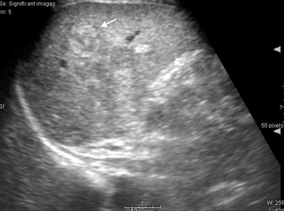
Fig. 2 A round hyperechoic lesion with a peripheral halo (arrow) was detected on ultrasonography.
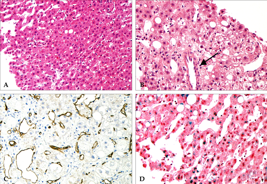
Fig. 3 Liver biopsy of the hypervascular hyperplastic nodule. The nodule shows slightly increased cellularity without cellular and structural atypia (A), unpaired arteries (arrow) (B), sinusoidal expression of CD34 (C) and focal iron deposition (D) (A; Hematoxylin-Eosin, × 100, B; Hematoxycin-Eosin, × 200, C; Labeled with Streptavidin Biotin, × 200, D; Prussian blue, × 200).
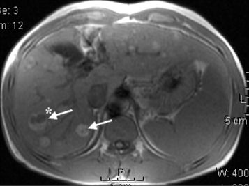
Fig. 4 Numerous nodules with a high signal on T1 weighted images, with a central low signal portion in some nodules (arrows), on the MRI. *The lesion was also detected on ultrasonography.
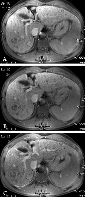
Fig. 5 Arterial phase enhancing nodules suggesting overt HCCs on the MRI (A: arterial phase, B: portal phase, C: delayed phase).

Fig. 6 Faint multiple nodular staining of both lobes of the liver in the early arterial phase on angiography.
Cited by 1 articles
-
A Case of Multiple Hypervascular Hyperplastic Liver Nodules in a Patient with No History of Alcohol Abuse or Chronic Liver Diseases
Byoung Joo Do, In Young Park, So Yon Rhee, Jin Kyung Song, Myoung Kuk Jang, Seong Jin Cho, Eun Sook Nam, Eun Joo Yun
Korean J Gastroenterol. 2015;65(5):321-325. doi: 10.4166/kjg.2015.65.5.321.
Reference
-
1. International Working Party. Terminology of nodular hepatocellular lesions. Hepatology. 1995. 22:983–993.2. Nagasue N, Akamizu H, Yukaya H, Yuuki I. Hepatocellular pseudotumor in the cirrhotic liver. Report of three cases. Cancer. 1984. 54:2487–2494.3. Nakashima O, Watanabe J, Tanaka M, Fukukura Y, Kojiro M, Kurohiji T, et al. Clinicopathological study of hyperplastic nodules in patient with chronic alcoholic liver disease. Acta Hepatol Jap. 1996. 37:704–713.4. Lepreux S, Laurent C, Balabaud C, Bioulac-Sage P. FNH-like nodules: Possible precursor lesions in patients with focal nodular hyperplasia (FNH). Comp Hepatol. 2003. 2:7.5. Kim SR, Maekawa Y, Imoto S, Sugano M, Kudo M. Hypervascular liver nodules in heavy drinkers of alcohol. Alcohol Clin Exp Res. 2004. 28:174S–180S.6. Nakashima O, Kurogi M, Yamaguchi R, Miyaaki H, Fujimoto M, Yano H, et al. Unique hypervascular nodules in alcoholic liver cirrhosis: identical to focal nodular hyperplasia-like nodules? J Hepatol. 2004. 41:992–998.7. Baron RL, Oliver JH, Dodd GD, Nalesnik M, Holbert BL, Carr B. Hepatocellular carcinoma: evaluation with biphasic, contrast-enhanced, helical CT. Radiology. 1996. 199:505–511.8. Matui O, Matui K, Kaneyama T, Yoshikawa J, Takashima T, Nakamura Y, et al. Benign and malignant nodules in cirrhotic livers: distinction based on blood supply. Radiology. 1991. 178:493–497.9. Peterson MS, Baron RL, Murakami T. Hepatic malignancies: usefulness of acquisition of multiple arterial and portal venous phase images at dynamic gadolinium-enhanced MR imaging. Radiology. 1996. 201:337–345.10. Suzuki M, Maeyama S, Takahashi H, Okuse N, Takahashi Y, Mizuno H, et al. Strategy for hepatic hyperplastic nodules in heavy drinkers. Alcohol Clin Exp Res. 2004. 28:153–158.11. Kondo F. Benign nodular hepatocellular lesions caused by abnormal hepatic circulation: etiological analysis and introduction of a new concept. J Gastroenterol Hepatol. 2001. 16:1319–1328.
- Full Text Links
-
- Actions
-
Cited
- CITED
-
- Close
- Share
-
- Similar articles
-
- A case of hypervascular hyperplastic nodules mimicking hepatocellular carcinoma in alcoholic liver cirrhosis
- A Case of Multiple Hypervascular Hyperplastic Liver Nodules in a Patient with No History of Alcohol Abuse or Chronic Liver Diseases
- Hypervascular Hyperplastic Nodules Appearing in Chronic Alcoholic Liver Disease: Benign Intrahepatic Nodules Mimicking Hepatocellular Carcinoma
- Focal nodular hyperplasia-like nodules in a young man with congenital liver cirrhosis
- Comparison of bacterial infection rate between patients with alcoholic liver cirrhosis and viral liver cirrhosis
