Yonsei Med J.
2013 Jan;54(1):41-47. 10.3349/ymj.2013.54.1.41.
Relationship between Angiographic Late Loss and 5-Year Clinical Outcome after Drug-Eluting Stent Implantation
- Affiliations
-
- 1Division of Cardiology, Severance Cardiovascular Hospital, Yonsei University College of Medicine, Seoul, Korea. mkhong61@yuhs.ac
- 2Severance Biomedical Science Institute, Yonsei University College of Medicine, Seoul, Korea.
- KMID: 1776916
- DOI: http://doi.org/10.3349/ymj.2013.54.1.41
Abstract
- PURPOSE
Currently, insufficient data exist to evaluate the relationship between angiographic late loss (LL) and long-term clinical outcome after drug-eluting stent (DES) implantation. In this study, we hypothesized that angiographic LL between 0.3 and 0.6 mm correlate with favorable long-term clinical outcomes.
MATERIALS AND METHODS
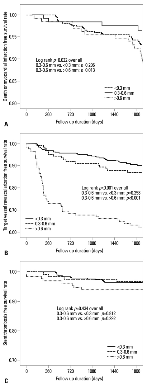
Patients were enrolled in the present study if they had undergone both DES implantation in single coronary vessel and a subsequent follow-up angiogram (n=634). These individuals were then subdivided into three groups based on their relative angiographic LL: group I (angiographic LL <0.3 mm, n=378), group II (angiographic LL between 0.3 and 0.6 mm, n=124), and group III (angiographic LL >0.6 mm, n=134). During a 5-year follow-up period, all subjects were tracked for critical events, defined as any cause of death or myocardial infarction, which were then compared among the three groups.
RESULTS
Mean follow-up duration was 63.0+/-10.0 months. Critical events occurred in 25 subjects in group I (6.6%), 5 in group II (4.0%), and 17 in group III (12.7%), (p=0.020; group I vs. group II, p=0.293; group II vs. group III, p=0.013). In a subsequent multivariate logistic regression analysis, chronic renal failure [odds ratio (OR)=3.29, 95% confidence interval (CI): 1.48-7.31, p=0.003] and long lesion length, defined as lesion length >28 mm (OR=1.88, 95% CI: 1.02-3.46, p=0.042) were independent predictors of long-term critical events.
CONCLUSION
This retrospective analysis fails to demonstrate that post-DES implantation angiographic LL between 0.3 and 0.6 mm is protective against future critical events.
Keyword
MeSH Terms
-
Adult
Aged
Angiography/*methods
Coronary Artery Disease/*surgery
Coronary Vessels/surgery
*Drug-Eluting Stents
Female
Follow-Up Studies
Humans
Kidney Failure, Chronic/complications
Male
Middle Aged
Multivariate Analysis
Odds Ratio
Percutaneous Coronary Intervention/methods
Prosthesis Failure
Retrospective Studies
Time Factors
Treatment Outcome
Figure
Reference
-
1. Tu JV, Bowen J, Chiu M, Ko DT, Austin PC, He Y, et al. Effectiveness and safety of drug-eluting stents in Ontario. N Engl J Med. 2007. 357:1393–1402.
Article2. Stone GW, Moses JW, Ellis SG, Schofer J, Dawkins KD, Morice MC, et al. Safety and efficacy of sirolimus- and paclitaxel-eluting coronary stents. N Engl J Med. 2007. 356:998–1008.
Article3. Daemen J, Wenaweser P, Tsuchida K, Abrecht L, Vaina S, Morger C, et al. Early and late coronary stent thrombosis of sirolimus-eluting and paclitaxel-eluting stents in routine clinical practice: data from a large two-institutional cohort study. Lancet. 2007. 369:667–678.
Article4. Iakovou I, Schmidt T, Bonizzoni E, Ge L, Sangiorgi GM, Stankovic G, et al. Incidence, predictors, and outcome of thrombosis after successful implantation of drug-eluting stents. JAMA. 2005. 293:2126–2130.
Article5. Finn AV, Joner M, Nakazawa G, Kolodgie F, Newell J, John MC, et al. Pathological correlates of late drug-eluting stent thrombosis: strut coverage as a marker of endothelialization. Circulation. 2007. 115:2435–2441.
Article6. Kim BK, Kim JS, Ko YG, Choi D, Jang Y, Hong MK. Correlation of angiographic late loss with neointimal coverage of drug-eluting stent struts on follow-up optical coherence tomography. Int J Cardiovasc Imaging. 2012. 28:1289–1297.
Article7. Kim U, Kim JS, Kim JS, Lee JM, Son JW, Kim J, et al. The initial extent of malapposition in ST-elevation myocardial infarction treated with drug-eluting stent: the usefulness of optical coherence tomography. Yonsei Med J. 2010. 51:332–338.
Article8. Pocock SJ, Lansky AJ, Mehran R, Popma JJ, Fahy MP, Na Y, et al. Angiographic surrogate end points in drug-eluting stent trials: a systematic evaluation based on individual patient data from 11 randomized, controlled trials. J Am Coll Cardiol. 2008. 51:23–32.9. Cutlip DE, Windecker S, Mehran R, Boam A, Cohen DJ, van Es GA, et al. Clinical end points in coronary stent trials: a case for standardized definitions. Circulation. 2007. 115:2344–2351.10. Mauri L, Orav EJ, O'Malley AJ, Moses JW, Leon MB, Holmes DR Jr, et al. Relationship of late loss in lumen diameter to coronary restenosis in sirolimus-eluting stents. Circulation. 2005. 111:321–327.
Article11. Brener SJ, Prasad AJ, Khan Z, Sacchi TJ. The relationship between late lumen loss and restenosis among various drug-eluting stents: a systematic review and meta-regression analysis of randomized clinical trials. Atherosclerosis. 2011. 214:158–162.
Article12. Carter AJ, Aggarwal M, Kopia GA, Tio F, Tsao PS, Kolata R, et al. Long-term effects of polymer-based, slow-release, sirolimus-eluting stents in a porcine coronary model. Cardiovasc Res. 2004. 63:617–624.
Article13. Farb A, Heller PF, Shroff S, Cheng L, Kolodgie FD, Carter AJ, et al. Pathological analysis of local delivery of paclitaxel via a polymer-coated stent. Circulation. 2001. 104:473–479.
Article14. Nakazawa G, Vorpahl M, Finn AV, Narula J, Virmani R. One step forward and two steps back with drug-eluting-stents: from preventing restenosis to causing late thrombosis and nouveau atherosclerosis. JACC Cardiovasc Imaging. 2009. 2:625–628.15. Nakazawa G, Otsuka F, Nakano M, Vorpahl M, Yazdani SK, Ladich E, et al. The pathology of neoatherosclerosis in human coronary implants bare-metal and drug-eluting stents. J Am Coll Cardiol. 2011. 57:1314–1322.16. Ko YG, Kim DM, Cho JM, Choi SY, Yoon JH, Kim JS, et al. Optical coherence tomography findings of very late stent thrombosis after drug-eluting stent implantation. Int J Cardiovasc Imaging. 2012. 28:715–723.
Article17. Takano M, Yamamoto M, Murakami D, Inami S, Okamatsu K, Seimiya K, et al. Lack of association between large angiographic late loss and low risk of in-stent thrombus: angioscopic comparison between paclitaxel- and sirolimus-eluting stents. Circ Cardiovasc Interv. 2008. 1:20–27.
Article18. Rivero F, Moreno R, Barreales L, Galeote G, Sánchez-Recalde A, Calvo L, et al. Lower levels of in-stent late loss are not associated with the risk of stent thrombosis in patients receiving drug-eluting stents. EuroIntervention. 2008. 4:124–132.
Article
- Full Text Links
- Actions
-
Cited
- CITED
-
- Close
- Share
- Similar articles
-
- Very Late Stent Thrombosis Related to Fracture of a Sirolimus-Eluting Stent
- Late Stent Thrombosis Associated with Late Stent Malapposition after Drug-Eluting Stenting: A Case Report
- A Case of Stent Thrombosis Occurred at 5 Years after Sirolimus-Eluting Stent Implantation
- Systemic drug therapy and restenosis after drug-eluting stent implantation
- Stent Thrombosis in the Era of the Drug-Eluting Stent


