A Case of Primary Biliary Malignant Lymphoma Mimicking Klatskin Tumor
- Affiliations
-
- 1Department of Internal Medicine, Inje University College of Medicine, Pusan Paik Hospital, Busan, Korea. cwj1225@naver.com
- KMID: 1775957
- DOI: http://doi.org/10.4166/kjg.2009.54.3.191
Abstract
- Primary non-Hodgkin's lymphoma of the extrahepatic bile duct presenting as obstructive jaundice is extremely rare. A 60-year-old man was admitted due to suddenly developed jaundice. Computerized tomography and endoscopic retrograde cholangiopancreatography showed a tumor at the proximal common hepatic duct. These clinical and radiologic findings resembled those of Klatskin tumor. The resection of the common hepatic duct tumor, lymph node dissection, and Roux-en-Y hepaticojejunostomy were carried out. There was no regional lymph node metastasis and no residual tumor at the resection margins. Histology and immunohistochemistry of the resected specimen confirmed a diffuse large B-cell malignant lymphoma involving the common hepatic duct. The patient is scheduled to receive adjuvant chemotherapy. In summary, primary non-Hodgkin's lymphoma of the extrahepatic bile duct, despite its rarity, should be considered in the differential diagnosis of causes for obstructive jaundice. An accurate histopathologic diagnosis and surgical resection combined with chemotherapy may be the approach to offer a chance for cure.
Keyword
MeSH Terms
Figure
-
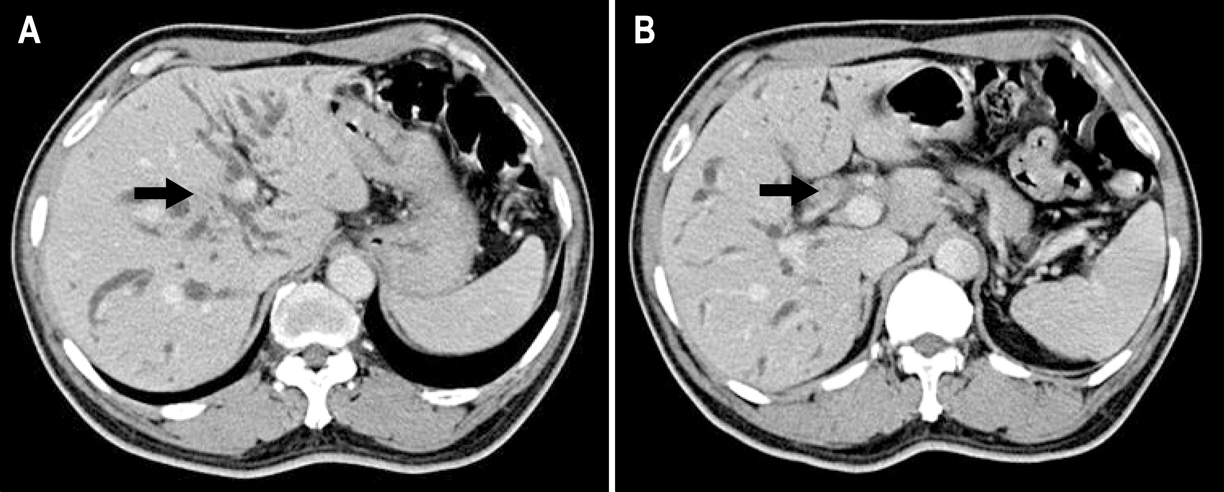
Fig. 1. Axial contrast-enhanced CT scans during the portal venous phase showed bile duct wall thickening of hyperattenuation from right secondary confluences (arrow in A) to the proximal common hepatic duct (arrow in B), and diffuse dilatation of both intrahepatic bile ducts.

Fig. 2. Magnetic resonance cholangiopancreatography (MRCP) showed stenosis of the main hepatic duct junction (arrow) with dilatation of intrahepatic bile ducts.
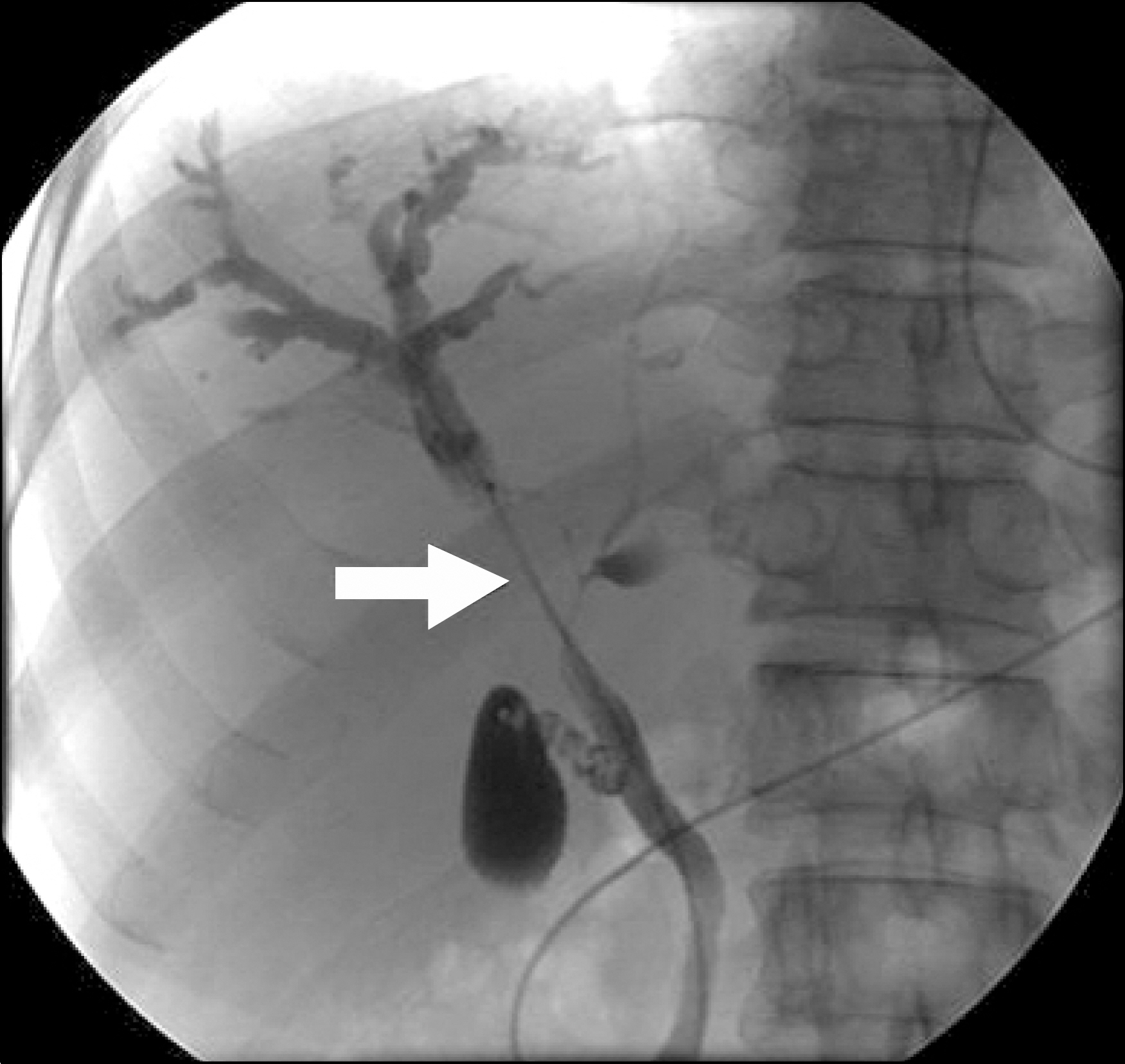
Fig. 3. Endoscopic nasobiliary drainage tube cholagiography revealed a long segment stricture at the hilar area with the visualization of right anterior superior segmental intrahepatic bile duct only, suggesting Klatskin tumor Bismuth type IIIa.
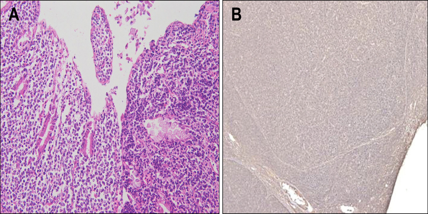
Fig. 4. (A) The resected specimen showed diffuse infiltrating neoplastic lymphoid cells at surrounding bile ducts and liver tissue (H&E stain, ×200), (B) Tumor cells were positive in immunohistochemical staining for the B cell-associated CD 20.
Cited by 1 articles
-
Primary Biliary Mucosa-associated Lymphoid Tissue Lymphoma Mimicking Hilar Cholangiocarcinoma
Seungha Hwang, Tae Jun Song, Seol So, Min Kyung Jeon, Eun Hye Oh, Byoung Soo Kwon, Sujong An, Myung-Hwan Kim
Korean J Gastroenterol. 2016;68(2):114-118. doi: 10.4166/kjg.2016.68.2.114.
Reference
-
1. Rosenberg SA, Diamond HD, Jaslowitz B, Craver LD. Lymphosarcoma: a review of 1269 cases. Medicine. 1961; 40:31–84.
Article2. Birrer MJ, Young RC. Differential diagnosis of jaundice in lymphoma patients. Semin Liver Dis. 1987; 7:269–277.
Article3. Lokich JJ, Kane RA, Harrison DA, McDermott WV. Biliary tract obstruction secondary to cancer: management guidelines and selected literature review. J Clin Oncol. 1987; 5:969–981.
Article4. Dudgeon DJ, Brower M. Primary chemotherapy for obstructive jaundice caused by intermediate-grade non-Hodgkin's lymphoma. Cancer. 1993; 71:2813–2816.5. Fidias P, Carey RW, Grossbard ML. Non-Hodgkin's lymphoma presenting with biliary tract obstruction. A discussion of seven patients and a review of the literature. Cancer. 1995; 75:1669–1677.
Article6. Ravindra KV, Stringer MD, Prasad KR, Kinsey SE, Lodge JP. Non-Hodgkin lymphoma presenting with obstructive jaundice. Br J Surg. 2003; 90:845–849.
Article7. Nguyen GK. Primary extranodal non-Hodgkin's lymphoma of the extrahepatic bile ducts. Cancer. 1982; 50:2218–2222.8. Kim SW, Kang SB, Kim CW, Yoon YB, Park YH. Primary lymphoma of the extrahepatic bile duct. Korean J Gastroenterol. 1997; 30:420–424.9. Joo YE, Park CH, Lee WS, et al. Primary non-Hodgkin's lymphoma of the common bile duct presenting as obstructive jaundice. J Gastroenterol. 2004; 39:692–696.
Article10. Levitan R, Diamond HD, Craver LF. Jaundice in Hodgkin's disease. Am J Med. 1961; 30:99–111.
Article11. Feller E, Schiffman FJ. Extrahepatic biliary obstruction by lymphoma. Arch Surg. 1990; 125:1507–1509.
Article12. Radhakrishnan S, Nakib BA, Liddawi HA, Ruwaih AA. Primary gastrointestinal lymphoma complicated by common bile duct obstruction: report of two cases. Am J Gastroenterol. 1986; 81:691–694.13. Ko YH, Kim CW, Park CS, et al. REAL classification of malignant lymphomas in the Republic of Korea: incidence of recently recognized entities and changes in clinicopathologic features. Hematolymphoreticular Study Group of the Korean Society of Pathologists. Revised European-American lymphoma. Cancer. 1998; 83:806–812.14. Jang EJ, Kim JS, Kim CW, et al. Clinical features and prognostic factors of primary intestinal lymphoma according to the cell lineage. Korean J Gastroenterol. 2002; 40:32–40.15. Kohno S, Ohshima K, Yoneda S, Kodama T, Shirakusa T, Kikuchi M. Clinicopathological analysis of 143 primary malignant lymphomas in the small and large intestines based on the new WHO classification. Histopathology. 2003; 43:135–143.
Article16. Shin ES, Yu CS, Huh JR, et al. Primary intestinal lymphoma. J Korean Surg Soc. 2003; 65:113–118.17. Takehara T, Matsuda H, Naitou M, et al. A case report of primary extranodal non-Hodgkin's lymphoma of the extrahepatic bile duct. Acta Hepatol Jpn. 1989; 88:247–252.
Article18. Tzanakakis GN, Vezeridis MP, Jackson BT, Rodil JV, McCully KS. Primary extranodal non-Hodgkin's lymphoma of the extrahepatic biliary tract. R I Med J. 1990; 73:483–486.19. Andre SB, Farias AQ, Bittencourt PL, et al. Primary extranodal non-Hodgkin's lymphoma of the extrahepatic bile duct mimicking klatskin tumor. Rev Hosp Clin Fac Med Sao Paulo. 1996; 51:192–194.
- Full Text Links
-
- Actions
-
Cited
- CITED
-
- Close
- Share
-
- Similar articles
-
- Primary Biliary Mucosa-associated Lymphoid Tissue Lymphoma Mimicking Hilar Cholangiocarcinoma
- Proximal Stent Migration of Fully Covered Self-Expandable Metal Stent following Side-by-Side Deployment in the Patient with Klatskin Tumor: A Case Report
- Mucosa-associated Lymphoid Tissue Lymphoma-mimicking Primary Gastrointestinal Small Lymphocytic Lymphoma
- A Case of Primary Malignant Lymphoma of Trachea
- Typical and Atypical Imaging Features of Malignant Lymphoma in the Abdomen and Mimicking Diseases
