Korean J Gastroenterol.
2010 Mar;55(3):154-161. 10.4166/kjg.2010.55.3.154.
Clinical Approach to Incidental Pancreatic Cystic Lesions
- Affiliations
-
- 1Department of Internal Medicine, Seoul National University College of Medicine, Seoul National University Bundang Hospital, Seongnam, Korea. gidoctor@snubh.org
- KMID: 1775896
- DOI: http://doi.org/10.4166/kjg.2010.55.3.154
Abstract
- Cystic lesions of the pancreas are being incidentally recognized with increasing frequency and become a common finding in clinical practice. Despite of recent remarkable advances of radiological and endoscopic assessment and a better understanding of natural history of certain subgroups of cystic lesions, differentiating among lesions and making an optimal management plan is still challenging. A multimodal approach should be performed to evaluate incidentally detected cystic lesions. Emerging evidence supports selective nonoperative management for the majority of patients with cystic lesions, but, for those in whom a suspicion of malignancy remains, surgery is indicated. Concerning long-term follow-up, there is limited data to support the ideal modality, intensity, and duration. Therefore, evidence-based guidelines for the diagnosis, management, and follow-up of cystic lesions of the pancreas should be established.
Keyword
MeSH Terms
-
Cystadenocarcinoma, Mucinous/diagnosis/epidemiology/therapy
Cystadenocarcinoma, Papillary/diagnosis/epidemiology/therapy
Cystadenocarcinoma, Serous/diagnosis/epidemiology/therapy
Humans
Incidence
Incidental Findings
Pancreatic Cyst/*diagnosis/epidemiology/therapy
Pancreatic Neoplasms/*diagnosis/epidemiology/therapy
Tomography, X-Ray Computed
Tumor Markers, Biological/blood
Figure
-
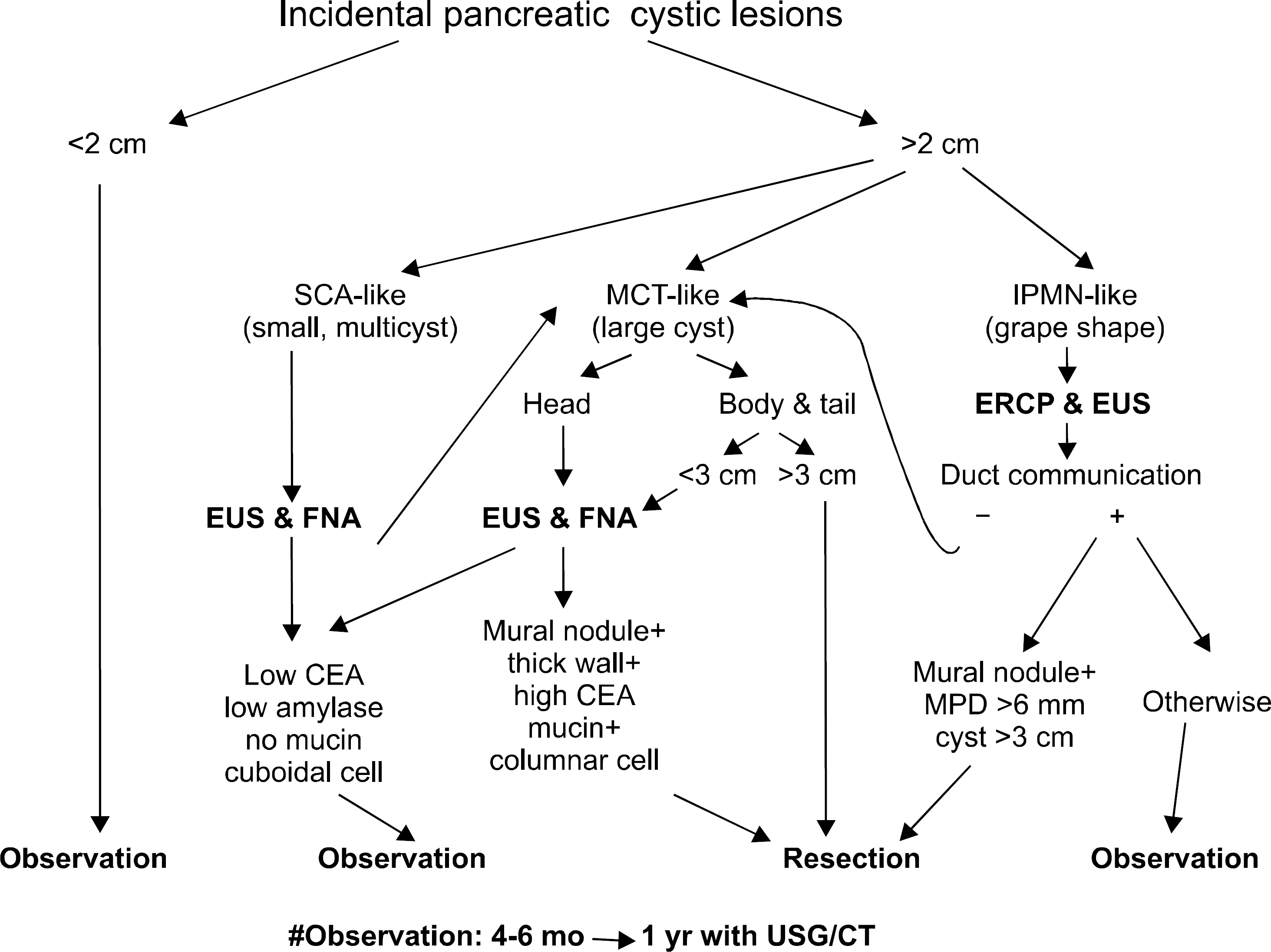
Fig. 1. Clinical approch to pancreatic cystic lesions. SCA, serous cystadenoma; MCT, mucinous cystic tumor; IPMN, intraductal papillary mucinous neoplasm; ERCP, endoscopic retrograde cholangiopancreatography; EUS, endoscopic ultrasonography; FNA, fine needle aspiration; MPD, main pancreatic duct; USG, ultrasonography; CT, computed tomography.
Reference
-
1. Edirimanne S, Connor SJ. Incidental pancreatic cystic lesions. World J Surg. 2008; 32:2028–2037.
Article2. Yoon WJ, Lee JK, Lee KH, Ryu JK, Kim YT, Yoon YB. Cystic neoplasms of the exocrine pancreas: an update of a nationwide survey in Korea. Pancreas. 2008; 37:254–258.3. Balthazar EJ, Chako AC. Computed tomography of pancreatic masses. Am J Gastroenterol. 1990; 85:343–349.4. Ferná ndez-del Castillo C, Targarona J, Thayer SP, Rattner DW, Brugge WR, Warshaw AL. Incidental pancreatic cysts: clinicopathologic characteristics and comparison with sympto-matic patients. Arch Surg. 2003; 138:427–433.5. Goh BK, Tan YM, Cheow PC, et al. Cystic lesions of the pancreas: an appraisal of an aggressive resectional policy adopted at a single institution during 15 years. Am J Surg. 2006; 192:148–154.
Article6. Allen PJ, D'Angelica M, Gonen M, et al. A selective approach to the resection of cystic lesions of the pancreas: results from 539 consecutive patients. Ann Surg. 2006; 244:572–582.
Article7. Lee SH, Shin CM, Park JK, et al. Outcomes of cystic lesions in the pancreas after extended follow-up. Dig Dis Sci. 2007; 52:2653–2659.
Article8. Salvia R, Crippa S, Falconi M, et al. Branch-duct intraductal papillary mucinous neoplasms of the pancreas: to operate or not to operate? Gut. 2007; 56:1086–1090.
Article9. Spinelli KS, Fromwiller TE, Daniel RA, et al. Cystic pancreatic neoplasms: observe or operate. Ann Surg. 2004; 239:651–657.10. Walsh RM, Vogt DP, Henderson JM, et al. Natural history of indeterminate pancreatic cysts. Surgery. 2005; 138:665–670.
Article11. Kimura W, Nagai H, Kuroda A, Muto T, Esaki Y. Analysis of small cystic lesions of the pancreas. Int J Pancreatol. 1995; 18:197–206.
Article12. Yoda Y, Suzuki Y, Yamada K, et al. A study of mass survey for pancreatic tumor using ultrasonography. J Gastroenterol Mass Surv. 1992; 96:50–55.13. Oh HC, Kim MH, Hwang CY, et al. Cystic lesions of the pancreas: challenging issues in clinical practice. Am J Gastroenterol. 2008; 103:229–239.
Article14. Le Borgne J, de Calan L, Partensky C. Cystadenomas and cystadenocarcinomas of the pancreas: a multiinstitutional retrospective study of 398 cases. French Surgical Association. Ann Surg. 1999; 230:152–161.15. Planner AC, Anderson EM, Slater A, Phillips-Hughes J, Bungay HK, Betts M. An evidence-based review for the management of cystic pancreatic lesions. Clin Radiol. 2007; 62:930–937.
Article16. Curry CA, Eng J, Horton KM, et al. CT of primary cystic pancreatic neoplasms: can CT be used for patient triage and treatment? Am J Roentgenol. 2000; 175:99–103.17. Box JC, Douglas HO. Management of cystic neoplasms of the pancreas. Am Surg. 2000; 66:495–501.18. Ferná ndez-del Castillo C, Warshaw AL. Current management of cystic neoplasms of the pancreas. Adv Surg. 2000; 34:237–248.19. Tseng JF, Warshaw AL, Sahani DV, Lauwers GY, Rattner DW, Fernandez-del Castillo C. Serous cystadenoma of the pancreas: tumor growth rates and recommendations for treatment. Ann Surg. 2005; 242:413–419.20. Balci NC, Semelka RC. Radiologic features of cystic, endocrine and other pancreatic neoplasms. Eur J Radiol. 2001; 38:113–119.
Article21. Buetow PC, Rao P, Thompson LD. From the Archives of the AFIP. Mucinous cystic neoplasms of the pancreas: radio-logic-pathologic correlation. Radiographics. 1998; 18:433–449.
Article22. Tanaka M, Chari S, Adsay V, et al. International consensus guidelines for management of intraductal papillary mucinous neoplasms and mucinous cystic neoplasms of the pancreas. Pancreatology. 2006; 6:17–32.
Article23. Ohashi K, Maruyama Y. Four cases of mucin producing cancer of the pancreas on specific findings of the ampulla of Vater. Prog Dig Endosc. 1982; 20:348–351.24. Salvia R, Ferná ndez-del Castillo C, Bassi C, et al. Main-duct intraductal papillary mucinous neoplasms of the pancreas: clinical predictors of malignancy and longterm survival following resection. Ann Surg. 2004; 239:678–685.25. Goh BK, Tan YM, Thng CH, et al. How useful are clinical, biochemical, and cross-sectional imaging features in predicting potentially malignant or malignant cystic lesions of the pancreas? Results from a single institution experience with 220 surgically treated patients. J Am Coll Surg. 2008; 206:17–27.
Article26. Friedrich CA. Von Hippel-Lindau syndrome. A pleomorphic condition. Cancer. 1999; 86:2478–2482.27. Sperti C, Pasquali C, Guolo P, Polverosi R, Liessi G, Pedrazzoli S. Serum tumor markers and cyst fluid analysis are useful for the diagnosis of pancreatic cystic tumors. Cancer. 1996; 78:237–243.
Article28. Ferná ndez-del Castillo C, Alsfasser G, Targarona J, Brugge WR, Warshaw AL. Serum CA 19-9 in the management of cystic lesions of the pancreas. Pancreas. 2006; 32:220.29. Ramage JK, Davies AH, Ardill J, et al. Guidelines for the management of gastroenteropancreatic neuroendocrine (including carcinoid) tumours. Gut. 2005; 54(suppl 4):iv1–16.
Article30. Johnson CD, Stephens DH, Charboneau JW, Carpenter HA, Welch TJ. Cystic pancreatic tumors: CT and sonographic assessment. AJR Am J Roentgenol. 1988; 151:1133–1138.
Article31. Procacci C, Biasiutti C, Carbognin G, et al. Characterization of cystic tumors of the pancreas: CT accuracy. J Comput Assist Tomogr. 1999; 23:906–912.
Article32. Walsh RM, Henderson JM, Vogt DP, et al. Prospective preoperative determination of mucinous pancreatic cystic neoplasms. Surgery. 2002; 132:628–633.
Article33. Cohen-Scali F, Vilgrain V, Brancatelli G, et al. Discrimination of unilocular macrocystic serous cystadenoma from pancreatic pseudocyst and mucinous cystadenoma with CT: initial observations. Radiology. 2003; 228:727–733.
Article34. Bassi C, Salvia R, Molinari E, Biasutti C, Falconi M, Pederzoli P. Management of 100 consecutive cases of pancreatic serous cystadenoma: wait for symptoms and see at imaging or vice versa? World J Surg. 2003; 27:319–323.
Article35. Gerke H, Jaffe TA, Mitchell RM, et al. Endoscopic ultrasound and computer tomography are inaccurate methods of classifying cystic pancreatic lesions. Dig Liver Dis. 2006; 38:39–44.
Article36. Fukukura Y, Fujiyoshi F, Hamada H, et al. Intraductal papillary mucinous tumors of the pancreas. Comparison of helical CT and MR imaging. Acta Radiol. 2003; 44:464–471.
Article37. Yamao K, Nakamura T, Suzuki T, et al. Endoscopic diagnosis and staging of mucinous cystic neoplasms and intraductal papillary-mucinous tumors. J Hepatobiliary Pancreat Surg. 2003; 10:142–146.
Article38. Lim SJ, Alasadi R, Wayne JD, et al. Preoperative evaluation of pancreatic cystic lesions: cost-benefit analysis and pro-posed management algorithm. Surgery. 2005; 138:672–679.
Article39. Sugiyama M, Atomi Y, Hachiya J. Intraductal papillary tumors of the pancreas: evaluation with magnetic resonance cholangiopancreatography. Am J Gastroenterol. 1998; 93:156–159.
Article40. Brugge WR, Lewandrowski K, Lee-Lewandrowski E, et al. Diagnosis of pancreatic cystic neoplasms: a report of the coperative pancreatic cyst study. Gastroenterology. 2004; 126:1330–1336.41. van der Waaij LA, van Dullemen HM, Porte RJ. Cyst fluid analysis in the differential diagnosis of pancreatic cystic lesions: a pooled analysis. Gastrointest Endosc. 2005; 62:383–389.
Article42. Linder JD, Geenen JE, Catalano MF. Cyst fluid analysis obtained by EUS-guided FNA in the evaluation of discrete cystic neoplasms of the pancreas: a prospective single-center experience. Gastrointest Endosc. 2006; 64:697–702.
Article43. Sperti C, Pasquali C, Decet G, Chierichetti F, Liessi G, Pedrazzoli S. F-18-fluorodeoxyglucose positron emission tomography in differentiating malignant from benign pancreatic cysts: a prospective study. J Gastrointest Surg. 2005; 9:22–28.
Article44. Mansour JC, Schwartz L, Pandit-Taskar N, et al. The utility of F-18 fluorodeoxyglucose whole body PET imaging for de-termining malignancy in cystic lesions of the pancreas. J Gastrointest Surg. 2006; 10:1354–1360.
Article45. Lahav M, Maor Y, Avidan B, Novis B, Bar-Meir S. Nonsurgical management of asymptomatic incidental pancreatic cysts. Clin Gastroenterol Hepatol. 2007; 5:813–817.
Article46. Ahn DW, Lee SH, Kim YT, et al. Natural history and clinical outcome after extended longterm follow-up for cystic lesions in the pancreas. Korean J Gastroenterol. 2009; 54:S299.47. Pelaez-Luna M, Chari ST, Smyrk TC, et al. Do consensus indications for resection in branch duct intraductal papillary mucinous neoplasm predict malignancy? A study of 147 patients. Am J Gastroenterol. 2007; 102:1759–1764.
Article48. Lee SH, Park JK, Woo SM, et al. Natural history of branch-duct type intraductal papillary mucinous neoplasms of the pancreas. Korean J Gastroenterol. 2007; 49:24–30.
Article49. Lee CJ, Scheiman J, Anderson MA, et al. Risk of malignancy in resected cystic tumors of the pancreas < or=3 cm in size: is it safe to observe asymptomatic patients? A multi-institutional report. J Gastrointest Surg. 2008; 12:234–242.50. Sahani DV, Kadavigere R, Saokar A, Fernandez-del Castillo C, Brugge WR, Hahn PF. Cystic pancreatic lesions: a simple imaging-based classification system for guiding management. Radiographics. 2005; 25:1471–1484.
Article51. Goh BK, Tan YM, Chung YF, et al. Pancreatic cysts: a pro-posed management algorithm based on current evidence. Am J Surg. 2007; 193:749–755.
Article52. Brugge WR, Lauwers GY, Sahani D, Fernandez-del Castillo C, Warshaw AL. Cystic neoplasms of the pancreas. N Engl J Med. 2004; 351:1218–1226.
Article
- Full Text Links
-
- Actions
-
Cited
- CITED
-
- Close
- Share
-
- Similar articles
-
- The Incidental Pancreatic Cyst: When to Worry About Cancer
- Recent Strategies for Differentiation and Management of Incidental Pancreatic Cystic Lesions
- Endoscopic Ultrasound-Guided Fine Needle Aspiration in Cystic Pancreatic Lesions
- Pathologic Features of Pancreatic Cystic Neoplasms
- Surgical Indications and Postsurgical Follow-up Strategy for Pancreatic Cystic Neoplasm
