Cholesterol Granuloma of the Tympanic Membrane Presenting as a Blue Eardrum
- Affiliations
-
- 1Department of Otolaryngology, Chonnam National University Medical School, Gwangju, Korea. chulsavio@hanmail.net
- 2Research Center for Resistant Cells, Chosun Medical School, Gwangju, Korea.
- KMID: 1758622
- DOI: http://doi.org/10.3349/ymj.2009.50.4.585
Abstract
- Intramembranous tympanic membrane cholesterol granuloma (CG) occurs infrequently. Here, the authors report a case of CG in the tympanic membrane presenting as a blue eardrum in the right ear. In addition, a pinhole perforation noted in the anterosuperior area revealed a brown discharge. High-resolution temporal bone CT showed a bulging mass shadow in the middle ear and a soft tissue dense lesion that filled both the epitympanum and mastoid cavity. Tympanomastoidectomy was performed under general anesthesia. New bone formation was confirmed in the mastoid antrum and epitympanum, and the epitympanum was blocked by new bone. The tympanic membrane revealed a round, brownish mass with a glistening surface and a severely thickened pars tensa. We herein report this case and review pertinent medical literature.
Keyword
MeSH Terms
Figure
-

Fig. 1 The pure tone audiogram showing that the hearing level in the right ear had scaled out at all frequencies.
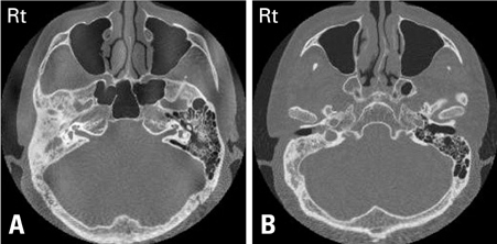
Fig. 2 High-resolution temporal bone CT findings of the middle ear and mastoid. (A) Temporal bone CT showing a dense soft tissue lesion filling both the epitympanum and mastoid cavity on the right side. New bone formation as a complication of childhood coalescent mastoiditis was noted. (B) Shows the bulging soft tissue in the middle ear.

Fig. 3 After elevation of the tympanomeatal flap, the inner surface of the tympanic membrane showed this round, brownish mass which had a glistening surface (arrow indicates) with severely thickened pars tensa at the posteroinferior quadrant.
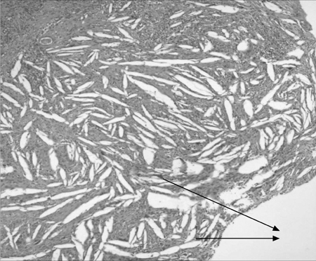
Fig. 4 Microscopic examination showing typical foreign body giant cells (arrows indicate) surrounding cholesterol crystals (Hematoxylin & Eosin stain, ×100)
Reference
-
1. Brackmann DE, Toh EH. Surgical management of petrous apex cholesterol granulomas. Otol Neurotol. 2002. 23:529–533.
Article2. Miglets AW, Booth JB. Cholesterol granuloma presenting as an isolated middle ear tumor. Laryngoscope. 1981. 91:410–415.
Article3. Hoffman RA. Cholesterol cyst manifesting a middle ear vascular tumor. Am J Otolaryngol. 1984. 5:68–70.4. Palva T, Lehto VP, Johnsson LG, Virtanen I, Mäkinen J. Large cholesterol granuloma cysts in the mastoid. Clinical and histopathologic findings. Arch Otolaryngol. 1985. 111:786–791.
Article5. Matt BH, Myer CM 3rd, Bellet PS. Cholesterol granuloma presenting the ear canal. Ann Otol Rhinol Laryngol. 1990. 99:672–673.6. da Costa SS, Paparella MM, Schachern PA, Yoon TH, Kimberley BP. Temporal bone histopathology in chronically infected ears with intact and perforated tympanic membranes. Laryngoscope. 1992. 102:1229–1236.
Article7. Haginomori S, Takamaki A, Ito K, Takenaka H, Kurisu Y, Tsuji M. Cholesterol granuloma in the tympanic membrane. Otol Neurotol. 2006. 27:1201–1202.
Article8. Nager GT, Vanderveen TS. Cholesterol granuloma involving the temporal bone. Ann Otol Rhinol Laryngol. 1976. 85:204–209.
Article9. Nager GT. Nager GT, Hyams VJ, editors. Cholesterol granuloma. Pathology of the ear and temporal bone. 1993. Baltimore: Williams & Wilkins;914–939.10. Jackler RK, Cho M. A new theory to explain the genesis of petrous apex cholesterol granuloma. Otol Neurotol. 2003. 24:96–106.
Article11. Miura M, Sando I, Orita Y, Hirsch BE. Histopathologic study of the temporal bones and Eustachian tubes of children with cholesterol granuloma. Ann Otol Rhinol Laryngol. 2002. 111:609–615.
Article12. Alaminos D, Gamboa J, Prades J. In : Sade J, editor. Cholesterol granuloma in the middle ear. Basic aspects of the Eustachian tube and middle ear disease. 1991. Conference on the Eustachian tube and middle ear disease; Genova: Kugler and Ghedini Publications;103–109.13. Leonetti JP, Shownkeen H, Marzo SJ. Incidental petrous apex findings on magnetic resonance imaging. Ear Nose Throat J. 2001. 80:200–202. 205–206.
Article14. Jaisinghani VJ, Paparella MM, Schachern PA, Le CT. Tympanic membrane/middle ear pathologic correlates in chronic otitis media. Laryngoscope. 1999. 109:712–716.15. Park K. Park KH, editor. Cholesterol granuloma of the middle ear. Middle ear diseases. 2002. Seoul: Academy'A;53–59.
- Full Text Links
-
- Actions
-
Cited
- CITED
-
- Close
- Share
-
- Similar articles
-
- Cholesterol Granuloma without Tympanic Membrane Perforation
- Middle Ear Cholesterol Granuloma without Tympanic Membrane Perforation
- Clinical Features of Cholesterol Granuloma in Temporal Bone
- Histologic findings of temporal bone in idiopathic blue eardrum
- Cholesterol Granuloma Presenting as Retroperitoneal Mass: A case report
