Castleman Disease of Hyaline Vascular Type in the Infrathyroidal Region: A Masquerader of Parathyroid Adenoma
- Affiliations
-
- 1Department of Radiology, Dongsan Medical Center, Keimyung University School of Medicine, Daegu, Korea. sklee@dsmc.or.kr
- 2Department of Pathology, Dongsan Medical Center, Keimyung University School of Medicine, Daegu, Korea.
- KMID: 1748451
- DOI: http://doi.org/10.3348/jksr.2013.68.3.201
Abstract
- Castleman disease of the infrathyroidal region is extremely rare. We report both CT and sonographic findings of a case of infrathyroidal paratracheal Castleman disease of hyaline vascular type, which masquerades parathyroid adenoma, in a 48-year-old woman. We further provide its histological findings at sonographically guided core-needle biopsy (US-CNB) and excisional biopsy. The lesion was ovoid with homogeneous intense enhancement on contrast-enhanced CT (CECT), and was homogeneous, markedly hypoechoic, and hypervascular on ultrasonography (US). Histological findings of the specimen obtained by US-CNB suggested lymphoproliferative lesion, and thus was inconclusive; those obtained by excisional biopsy were characteristics of Castleman disease of hyaline vascular type. Hyaline vascular type Castleman's disease should be included in the differential diagnosis of a mass of the infrathyroidal region with homogeneous intense enhancement on CECT, as well as with marked hypoechogenicity and hypervascularity on US. US-CNB may be of limited value in the histological diagnosis of this entity.
MeSH Terms
Figure
-
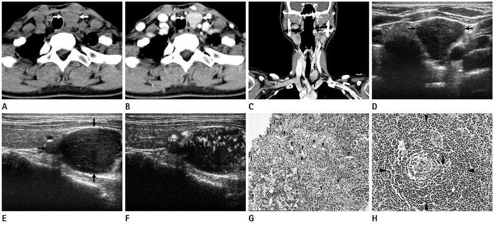
Fig. 1 Castleman disease of hyaline vascular type in the left infrathyroidal paratracheal region (level VI) in a 48-year-old woman. A. A non-enhanced CT image shows a well-demarcated, ovoid, slightly lobular, and homogeneous solid mass (arrows) with attenuation value similar to adjacent muscles. B, C. Contrast-enhanced axial and coronal reformatted CT images reveal an infrathyroidal paratracheal mass with homogeneous, intense enhancement (arrows). D, E. Transverse and longitudinal gray-scale sonographic images show a well-demarcated, homogeneous, and markedly hypoechoic solid mass (arrows) in the left infrathyroidal paratracheal region. F. A longitudinal power Doppler sonographic image reveals prominent vascularity in the central and peripheral portions of the mass. G. A photomicrograph of histological examination of the specimen obtained by sonographically guided core-needle biopsy demonstrates numerous small lymphocytes and markedly increased vascularity (arrowheads) without germinal center (hematoxylin-eosin, × 200), and immunohistochemical staining for CD20 reveals strong positivity for numerous small B cells (inset: CD20, × 200). H. A photomicrograph of the histological examination of the specimen obtained by surgical excision exhibits a germinal center pierced by a hyalinized blood vessel (arrow) ("lollipop" appearance) and cuffed by the characteristically expanded mantle zone composed of small lymphocytes arranged in a concentric onionskin pattern (arrowheads), which are characteristic of Castleman disease of hyaline vascular type (hematoxylin-eosin, × 400).
Reference
-
1. Cronin DM, Warnke RA. Castleman disease: an update on classification and the spectrum of associated lesions. Adv Anat Pathol. 2009. 16:236–246.2. Chen YF, Zhang WD, Sun CZ, Ouyang D, Chen WK, Luo RZ, et al. Clinical features and outcomes of head and neck castleman disease. J Oral Maxillofac Surg. 2012. 70:2466–2479.3. Lin CY, Chang YL. Castleman's disease in the head and neck region: Meta-analysis of reported cases in Taiwan and literature review. J Formos Med Assoc. 2010. 109:913–920.4. Song JJ, Jung MH, Woo JS, Chae SW, Hwang SJ, Lee HM. Castleman's disease of the head and neck. Eur Arch Otorhinolaryngol. 2006. 263:160–163.5. Keller AR, Hochholzer L, Castleman B. Hyaline-vascular and plasma-cell types of giant lymph node hyperplasia of the mediastinum and other locations. Cancer. 1972. 29:670–683.6. Tan TY, Pang KP, Goh HK, Teo EL, Abhilash B, Walford N. Castleman's disease of the neck: a description of four cases on contrast-enhanced CT. Br J Radiol. 2004. 77:253–256.7. Glazer M, Rao VM, Reiter D, McCue P. Isolated Castleman disease of the neck: MR findings. AJNR Am J Neuroradiol. 1995. 16:669–671.8. Mahmood N, Suresh HB, Swethadri GK, Hegde V, D'Souza V, D'Souza S. Ultrasound and Doppler findings in a rare case of Castleman's disease of the parotid. Dentomaxillofac Radiol. 2010. 39:54–56.9. Souza KC, Silva SJ, Salomão E, Silva AM, Faria PR, Queiroz LF, et al. Cervical Castleman's disease in childhood. J Oral Maxillofac Surg. 2008. 66:1067–1072.10. Huppert BJ, Reading CC. Parathyroid sonography: imaging and intervention. J Clin Ultrasound. 2007. 35:144–155.
