J Korean Soc Spine Surg.
2008 Sep;15(3):199-203. 10.4184/jkss.2008.15.3.199.
Perineural Cyst in Upper Lumbar Spine: A Case Report
- Affiliations
-
- 1Department of Orthopedic Surgery, Hanil General Hospital, Seoul, Korea. pooljang105@hanmail.net
- KMID: 1747205
- DOI: http://doi.org/10.4184/jkss.2008.15.3.199
Abstract
- Spinal perineural cyst rarely appears in the upper lumbar spinal region as compared it occurrence at other spinal region. These cysts seldom cause radiating pain and neurologic symptoms because the majority of these cysts involve the sacral portion of the spine. Spinal perineural cyst can be recognized incidentally on magnetic resonance imaging while differentiating other diseases that cause back pain. The differential diagnosis of symptoms is needed to rule out whether or not the intraspinal cystic mass is the actual cause of radiculopathy. We report here on a case of perineural cyst at a left neural foramen of L2-3 and the patient displayed neurologic manifestations. The patient had progressively aggravated low back pain and sciatica of two years duration. MRI well demonstrated neural compression of the left 2nd lumbar neural root. Relief of symptoms were achieved by performing posterior decompression (hemilaminectomy, fascectomy & decompression of the ligamentum flavum), excision of the cyst and posterolateral fusion of L2-3.
Keyword
MeSH Terms
Figure
-

Fig. 1. MR images show perineural cyst located on the Lt neural foramen of L2-3 level. Sagital & axial T2-weighted image: 1×1.2 ×0.9 cm sized mass with high signal in T2 images & Intraoperative surgical findings showing an enlarged Lt L2 nerve root compressing the dura
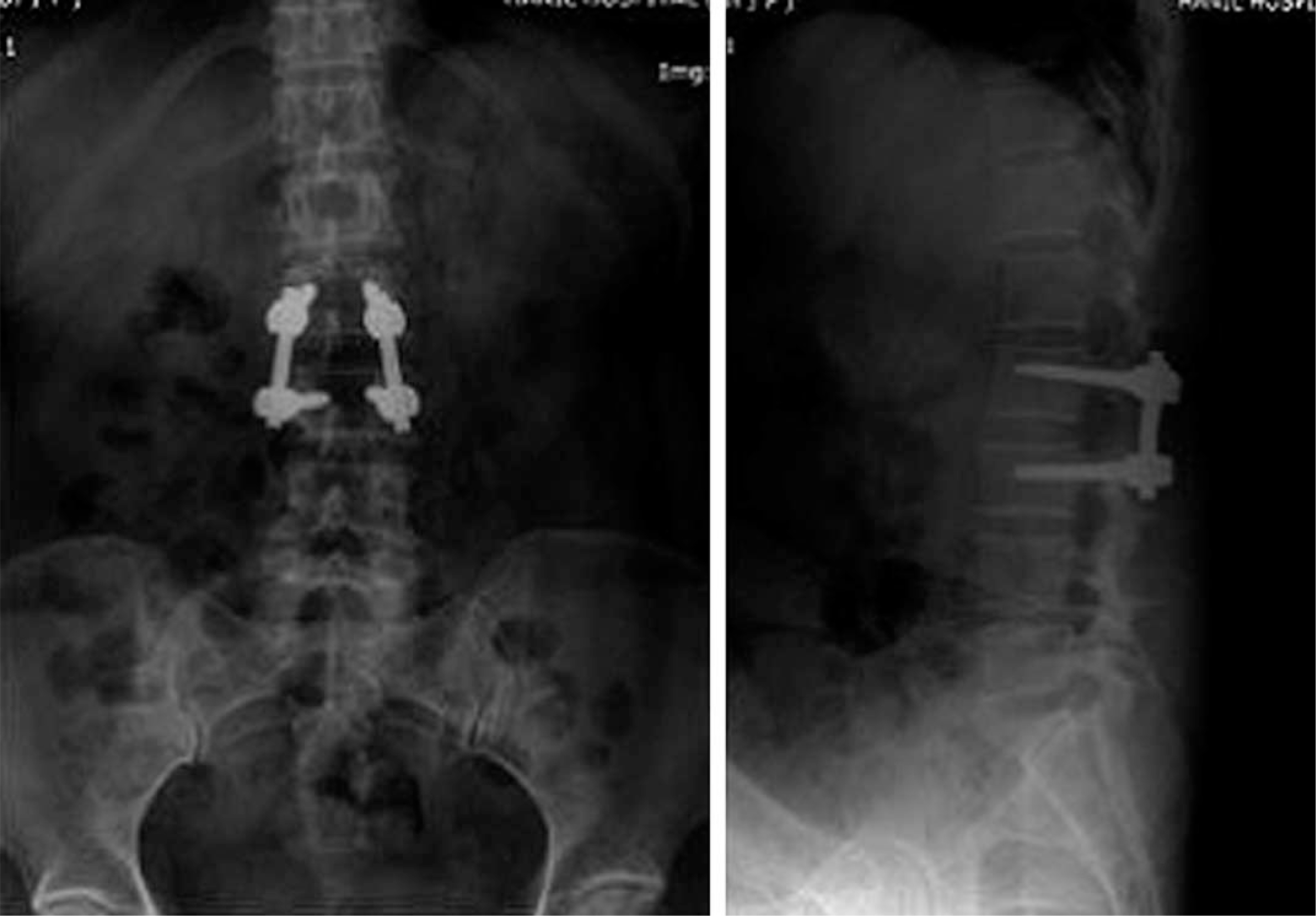
Fig. 2. Postoperative lumbar X-ray AP, lateral image showing fusion state between L2-3

Fig. 3. Postoperative sagittal & axial T2 image of taken 6 months showing complete dural decompression and no evidence of recurrence.

Fig. 4. Cytology finding showing clusted lymphocyte and methothelial cell in intracyst fluid
Reference
-
1). Tarlov IM. Cysts (Perineurial) of the sacral roots. Another cause (removal) of sciatic pain. JAMA. 1948; 138:740–744.2). Ziegler DK, Batnitzky S. Coccygodynia caused by perineural cyst. Neurology. 1984; 34:829–830.
Article3). Rodziewicz GS, Kaufman B, Spetzler RF. Diagnosis of Sacral Perineural Cysts by Nuclear Magnetic Resonance. Surg Neurol. 1984; 22:50–52.
Article4). Lee KS, Kim BS, Choi JC, Park BY, Cha JH. Sacral Perineural Cyst-report of 3 cases-. J Koreal Orthop Assoc. 1997; 32:1085–1089.5). Jain SK, Chopra S, Bagaria H, Mathur PP. Sacral perineural cyst presenting as chronic perineal pain: a case report. Neurol India. 2002; 50:514–515.6). Bassiouni H, Hunold A, Asgari S, Hubschen U, Konig HJ, Stolke D. Spinal intradural juxtamedullary cysts in the adult: surgical management and outcome. Neurosurgery. 2004; 55:1352–1359.
Article7). Edir BS, Leslie S, Leonard IK, Joseph G. CT character-ristics of sacral perineural cysts, Report of two cases. J Neurosurg. 1984; 61:596–598.8). McCrum C, William B. Spinal extradural arachnoid pouches. Report of two cases. J Neurosurg. 1982; 57:849–852.9). KS Lee, JC Choi, YJ Kim, U Jin, YJ Jung. Spinal meningeal cyst in low back patients. J Korean Orthop Assoc. 1998; 33:1600–1605.10). Benzel EC. Spine surgery. 2nd ed.Elsevier. Church Livingstone: 1124;2005.
- Full Text Links
-
- Actions
-
Cited
- CITED
-
- Close
- Share
-
- Similar articles
-
- Sacral Perineural Cyst with Lumbar Disc Herniation: A Case Report
- Cervical Perineural Cyst Masquerading as a Cervical Spinal Tumor
- Treatment of Cervical Perineural Cyst by the Transforaminal Epidural Steroid Injection
- Sacral Perineural Cyst: Another Cause of Sciatica
- Multiple Bilateral Thoracic Perineural Cysts: A Case Report
